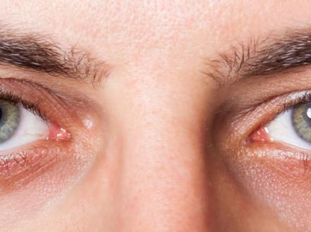
Allergic conjunctivitis is inflammation of the bulbar and palpebral conjunctiva caused by an IgE‑mediated hypersensitivity to environmental allergens (eg, pollens, animal dander, dust mites) or, in some forms, chronic T‑cell–mediated inflammation. It typically causes intense itching, tearing, redness, and chemosis (conjunctival edema). Diagnosis is clinical based on history and slit‑lamp examination; laboratory testing is rarely needed. Treatment centers on allergen avoidance, cold compresses, lubricants, and topical dual‑action antihistamine/mast‑cell stabilizer drops; short courses of topical corticosteroids are reserved for severe disease or corneal involvement.
Key Points
- Itching is the hallmark symptom of allergic conjunctivitis; pain and photophobia suggest alternate or complicated diagnoses.
- First‑line therapy for most patients is a topical dual‑action antihistamine/mast‑cell stabilizer (eg, olopatadine, ketotifen), plus allergen avoidance and cold compresses.
- Seasonal (SAC) and perennial (PAC) forms are common and usually mild; vernal (VKC) and atopic keratoconjunctivitis (AKC) are sight‑threatening variants requiring specialist care.
- Avoid contact lenses during active inflammation; contact lens–related giant papillary conjunctivitis (GPC) improves with discontinuation and hygiene changes.
- Topical corticosteroids are effective for severe flares but require intraocular pressure (IOP) monitoring due to risks of glaucoma, cataract, and infection reactivation.
- Consider allergen immunotherapy for patients with concomitant allergic rhinitis whose ocular symptoms recur each season.
Anatomy and Physiology
The conjunctiva is a thin, vascularized mucous membrane lining the posterior eyelids (palpebral conjunctiva) and reflecting onto the globe (bulbar conjunctiva). Goblet cells produce mucins critical to tear film stability. The tarsal conjunctiva harbors immune effector cells, including mast cells and eosinophils. The superior tarsal plate forms prominent papillae in allergic disease; limbal tissue can form Horner‑Trantas dots (eosinophil aggregates) in VKC.
Etiology
- Environmental aeroallergens: pollens (trees, grasses, weeds), house dust mites, animal dander, molds.
- Mechanical/foreign body–related: contact lenses (especially reusable soft lenses), exposed sutures, ocular prostheses (GPC).
- Atopic diathesis: association with atopic dermatitis, asthma, and allergic rhinitis (notably in AKC and VKC).
- Occupational/indoor triggers: feathers, chemicals, occupational dusts.
- Medication‑related conjunctival hypersensitivity or medicamentosa (eg, preservatives like benzalkonium chloride).
Pathophysiology
- Immediate (Type I) hypersensitivity: Allergen cross‑links IgE on conjunctival mast cells, releasing histamine and other mediators, causing vasodilation, itching via H1 receptors, and chemosis (SAC/PAC).
- Late‑phase and chronic inflammation: Recruitment of eosinophils, Th2 lymphocytes, and cytokines sustains inflammation, leading to papillary hypertrophy, mucus hypersecretion, and, in severe forms (VKC/AKC), corneal epithelial toxicity and shield ulcers.
- Mechanical microtrauma: Contact lens edge and deposits stimulate papillary hypertrophy (GPC).
Epidemiology
Allergic conjunctivitis is common in the United States and frequently coexists with allergic rhinitis. Prevalence estimates vary by method and region; population surveys suggest that a substantial proportion of individuals with seasonal allergies report ocular itching and redness each year. VKC is uncommon in temperate climates and primarily affects boys in late childhood; AKC typically presents in young adults with chronic atopic dermatitis [1][2].
Classification
- Seasonal allergic conjunctivitis (SAC): Intermittent, pollen‑driven flares (spring/summer/fall).
- Perennial allergic conjunctivitis (PAC): Year‑round, often due to dust mites, animal dander, molds; milder but chronic.
- Vernal keratoconjunctivitis (VKC): Chronic, seasonal exacerbations; large superior tarsal papillae, limbal inflammation, risk of shield ulcers; pediatric‑predominant.
- Atopic keratoconjunctivitis (AKC): Chronic, often bilateral, associated with atopic dermatitis; can cause cicatrization, keratopathy, and vision loss.
- Giant papillary conjunctivitis (GPC), also called contact lens–induced papillary conjunctivitis (CLPC): Papillary hypertrophy of the superior tarsal conjunctiva from contact lenses, exposed sutures, or prostheses.
Symptoms and Signs
Seasonal and Perennial Allergic Conjunctivitis (SAC/PAC)
- Symptoms: Intense itching, tearing (epiphora), burning, mild photophobia, watery or stringy mucus discharge, eyelid swelling. Often with allergic rhinitis (sneezing, nasal congestion).
- Signs: Conjunctival hyperemia, chemosis, mild eyelid edema, papillary reaction (small papillae) on tarsal conjunctiva; clear cornea.
Vernal Keratoconjunctivitis (VKC)
- Symptoms: Severe itching, photophobia, foreign‑body sensation, ropy mucus discharge; seasonal worsening.
- Signs: Giant “cobblestone” papillae on superior tarsal conjunctiva; limbal thickening with Horner‑Trantas dots; punctate keratopathy; risk of superior corneal shield ulcers and plaques.
Atopic Keratoconjunctivitis (AKC)
- Symptoms: Chronic itching, burning, photophobia, mucus discharge; often perennial with flares.
- Signs: Lid skin eczema with fissuring; tarsal papillae; conjunctival scarring; symblepharon in severe cases; punctate epithelial erosions; keratoconus association reported [2].
Giant Papillary Conjunctivitis (GPC/CLPC)
- Symptoms: Itching worse after lens removal, mucus discharge, decreased lens tolerance, lens awareness.
- Signs: Enlarged superior tarsal papillae, mucus strands, lens deposits; cornea usually clear unless coexisting keratitis.
Complications
- Corneal involvement: Punctate epithelial erosions, shield ulcers (VKC), microbial keratitis from eye rubbing or lens wear.
- Vision impairment: From corneal scarring, irregular astigmatism, or keratoconus progression [2].
- Medication adverse effects: Steroid‑induced ocular hypertension/glaucoma, posterior subcapsular cataract, herpetic keratitis reactivation; rebound hyperemia with chronic vasoconstrictor use.
- Contact lens complications: Intolerance, increased risk of infectious keratitis.
Diagnosis
Clinical evaluation
- History: Onset relative to allergen exposure or seasons, severity of itching, presence of rhinitis/asthma/atopic dermatitis, contact lens type and hygiene, prior response to drops, use of vasoconstrictors or preserved medications.
- Examination: Visual acuity; external and slit‑lamp exam for tarsal papillae, hyperemia, chemosis, limbal changes, Trantas dots; fluorescein staining to assess corneal epithelium; eyelid skin for dermatitis.
- Pain, marked photophobia, or reduced vision warrants careful corneal evaluation to exclude keratitis or uveitis.
Imaging and laboratory testing
- Not routinely required.
- Conjunctival cytology may show eosinophils; helpful in atypical or refractory cases.
- Allergen evaluation (skin‑prick or serum‑specific IgE) guided by history when considering allergen immunotherapy or when diagnosis is uncertain; coordinate with allergy specialists [3][4].
Differential diagnosis
| Entity | Distinguishing features |
|---|---|
| Viral conjunctivitis (adenoviral) | Follicles, preauricular lymphadenopathy, watery discharge, recent URI; highly contagious |
| Bacterial conjunctivitis | Mucopurulent discharge, eyelids stuck in morning, minimal itching; no seasonal pattern |
| Dry eye disease | Burning, fluctuating vision, worsens late day, positive staining of interpalpebral cornea; minimal itching |
| Blepharitis/meibomian gland dysfunction | Lid margin crusting, chalazia, morning irritation; improves with lid hygiene |
| Contact dermatitis of eyelids | Lid erythema/edema, pruritus, scaling; periocular exposure history |
| Drug‑induced conjunctivitis (medicamentosa) | Chronic redness on preserved drops (eg, glaucoma meds), inferior conjunctival changes; improves with discontinuation |
| Keratitis (bacterial, herpetic, Acanthamoeba) | Pain, photophobia, focal corneal infiltrate/ulcer, anterior chamber reaction; urgent evaluation needed |
| Anterior uveitis (iritis) | Photophobia, ciliary flush, cells/flare in anterior chamber; small pupil |
| Scleritis/episcleritis | Focal/diffuse deep redness; scleritis has severe pain and tenderness |
| Acute angle‑closure glaucoma | Severe pain, halos, mid‑dilated pupil, corneal edema, high IOP; emergency |
| Subconjunctival hemorrhage | Painless, sharply demarcated red patch; no discharge or itching |
Treatment
Medical management
Nonpharmacologic
- Allergen avoidance (keep windows closed during pollen peaks, shower after outdoor exposure, use high‑efficiency particulate air [HEPA] filtration).
- Cold compresses 10–15 minutes as needed; preservative‑free artificial tears 4–8 times/day to dilute allergens and mediators.
- Suspend contact lens wear during active inflammation; consider daily disposable lenses once quiet.
Topical antihistamine/mast‑cell stabilizer combinations (first‑line for SAC/PAC)
- Olopatadine 0.1% 1 drop bid or 0.2% qd.
- Ketotifen 0.025% (OTC) 1 drop bid.
- Alcaftadine 0.25% 1 drop qd.
- Epinastine 0.05% 1 drop bid.
- Bepotastine 1.5% 1 drop bid.
- Azelastine 0.05% 1 drop bid.
- Onset within minutes; continue through the allergen season for prevention [1][5].
Mast‑cell stabilizers (for prophylaxis; slower onset over days–weeks)
- Cromolyn sodium 4% 1 drop qid.
- Nedocromil 2% 1 drop bid.
- Lodoxamide 0.1% 1 drop qid.
Adjuncts
- Oral second‑generation antihistamines (cetirizine 10 mg PO qd, loratadine 10 mg PO qd, fexofenadine 180 mg PO qd) for patients with significant rhinitis; may worsen ocular dryness.
- Topical NSAIDs (eg, ketorolac 0.5% 1 drop qid) have limited additional benefit and may sting; not first‑line [1].
- Topical vasoconstrictor/antihistamine OTC combinations (eg, naphazoline/pheniramine) can reduce redness/itching short‑term; limit to brief use due to rebound hyperemia and risk of angle‑closure in susceptible eyes; avoid in narrow‑angle glaucoma [DailyMed/FDA label].
Topical corticosteroids (for severe flares, VKC/AKC with corneal involvement)
- Loteprednol etabonate 0.2–0.5% 1 drop qid (then taper).
- Fluorometholone 0.1% 1 drop qid (then taper).
- Use the lowest effective dose for the shortest duration; monitor IOP and lens status; exclude active herpetic keratitis [1][6].
Calcineurin inhibitors (steroid‑sparing; off‑label in the US for allergic indications)
- Cyclosporine ophthalmic emulsion 0.05% 1 drop bid for chronic VKC/AKC refractory to standard therapy (FDA‑approved for dry eye; off‑label here) [2].
- Tacrolimus 0.03% dermatologic ointment applied thinly to eyelid skin bid for lid eczema in AKC; avoid direct instillation unless compounded; off‑label [2].
Allergen immunotherapy
- Subcutaneous or sublingual allergen immunotherapy can reduce ocular symptoms in SAC/PAC with concomitant allergic rhinitis when targeted to confirmed allergens; coordinated with an allergist [3][4].
GPC‑specific measures
- Discontinue contact lens wear for 2–4 weeks; switch to daily disposables or rigid gas‑permeable lenses; improve lens hygiene and replace frequently.
- Treat inflammation with dual‑action drops; consider a short steroid course if severe.
Procedural and surgical management
- VKC shield ulcers with non‑adherent plaques: Debridement of plaque and placement of a bandage soft contact lens; topical antibiotic prophylaxis during epithelial healing.
- Recalcitrant VKC/AKC: Consider supratarsal corticosteroid injection in select cases under ophthalmologist supervision; reserved due to risk profile [2].
- Mechanical sources of GPC (exposed sutures): Remove or revise the irritant.
Special populations
- Pediatric: VKC occurs in school‑age boys; prioritize dual‑action drops, allergen avoidance, and careful steroid use with IOP monitoring. Educate to avoid eye rubbing to reduce keratopathy risk.
- Pregnancy/lactation: Prefer nonpharmacologic measures first. Topical dual‑action agents have low systemic absorption; use if benefits outweigh risks. Avoid prolonged use of topical vasoconstrictors. Coordinate with obstetric providers; choose oral agents with established safety if needed (eg, loratadine, cetirizine).
- Immunocompromised: Use steroids cautiously; rule out herpetic keratitis before initiating. Monitor closely for infection.
- Older adults: Oral antihistamines may exacerbate dry eye and can cause sedation; avoid first‑generation agents. Avoid vasoconstrictors in narrow‑angle glaucoma.
Prognosis
- SAC/PAC: Excellent with avoidance and topical therapy; symptoms typically remit when exposure ends.
- GPC: Improves with lens holiday and hygiene modifications; recurrence possible if risk factors persist.
- VKC: Often improves after adolescence; corneal complications can threaten vision without aggressive control.
- AKC: Chronic relapsing course into adulthood with risk of cicatrization and keratopathy; requires long‑term management and monitoring [1][2].
Prevention and Patient Counseling
- Start dual‑action drops 1–2 weeks before predictable pollen seasons (SAC).
- Minimize exposure: check pollen counts, keep windows closed, use air conditioning with high‑quality filters, shower after outdoor activities, and avoid rubbing eyes.
- Use preservative‑free artificial tears regularly during high‑exposure days.
- Avoid chronic use (>2 weeks) of OTC “redness relief” vasoconstrictor drops.
- Suspend contact lens wear during active symptoms; consider daily disposables thereafter.
- Adhere to follow‑up when using topical steroids for IOP checks.
When to Seek Immediate Care
- Sudden decrease in vision in either eye.
- Moderate to severe eye pain or significant light sensitivity.
- A white spot on the cornea or suspected corneal ulcer.
- Copious pus‑like discharge or eyelids matted shut throughout the day.
- Symptoms in a contact lens wearer that do not improve promptly after removing lenses.
- Use of steroid eye drops without medical supervision.
References
- American Academy of Ophthalmology Cornea/External Disease Panel. Conjunctivitis Preferred Practice Pattern. San Francisco, CA: AAO; 2023. https://www.aao.org/preferred-practice-pattern/conjunctivitis-ppp-2023
- EyeWiki (AAO). Allergic Conjunctivitis. https://eyewiki.aao.org/Allergic_Conjunctivitis
- MedlinePlus. Allergic Conjunctivitis. National Library of Medicine. https://medlineplus.gov/ency/article/001011.htm
- Wallace DV, et al. The diagnosis and management of rhinitis: An updated practice parameter. J Allergy Clin Immunol. 2008;122(2 Suppl):S1-S84. doi:10.1016/j.jaci.2008.06.003
- Calderon MA, et al. Topical antihistamines and mast cell stabilisers for seasonal and perennial allergic conjunctivitis. Cochrane Database Syst Rev. 2015;(6):CD009566. doi:10.1002/14651858.CD009566.pub2
- DailyMed. Loteprednol Etabonate Ophthalmic Suspension. U.S. National Library of Medicine. https://dailymed.nlm.nih.gov
Closing disclaimer: This article is for informational purposes and is not a substitute for professional medical advice, diagnosis, or treatment. Always consult a qualified eye care professional about your specific condition. If you have sudden vision loss, severe eye pain, or an eye injury, seek emergency care immediately.