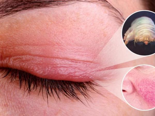
Demodex refers to overgrowth of commensal Demodex mites—primarily Demodex folliculorum and Demodex brevis—on the eyelids and eyelashes, causing chronic anterior blepharitis and meibomian gland dysfunction. Cardinal findings are cylindrical collarettes (sleeves) at the lash bases, lid margin inflammation, itching, foreign body sensation, and ocular surface irritation. Diagnosis is clinical at the slit lamp. Treatment includes lid hygiene, targeted acaricidal therapy (lotilaner ophthalmic solution 0.25%), and adjunctive measures for inflammation and associated rosacea/meibomian disease.
Key Points
- Demodex folliculorum infests lash follicles; Demodex brevis inhabits sebaceous/meibomian glands and is linked to meibomian gland dysfunction (MGD) and chalazia.
- Cylindrical “collarettes” at the lash base are the most specific sign of Demodex blepharitis.
- Diagnosis is clinical; lash epilation microscopy confirms mites but is seldom required in routine practice.
- FDA‑approved lotilaner ophthalmic solution 0.25% is dosed 1 drop in each eye bid for 6 weeks for Demodex blepharitis.
- Adjuncts include mechanical lid debridement, warm compresses, and management of ocular rosacea/MGD; tea tree oil derivatives are used off‑label and may irritate ocular tissues.
- Untreated disease can contribute to chronic blepharitis, recurrent chalazia, keratitis, and dry eye symptoms.
Anatomy and Physiology
- Eyelash follicles and associated sebaceous glands (glands of Zeis) line the anterior lid margin; meibomian glands in the tarsus secrete the lipid layer of the tear film.
- D. folliculorum resides in lash follicles and feeds on follicular debris and epithelial cells. D. brevis penetrates sebaceous and meibomian glands.
- The tear film’s lipid layer stabilizes the aqueous layer; meibomian gland dysfunction increases tear evaporation and ocular surface inflammation.
Etiology
- Overproliferation of Demodex mites on eyelid skin and adnexa.
- Predisposing factors: increasing age, ocular or facial rosacea, seborrheic dermatitis, chronic lid hygiene lapses, contact lens wear, and immunosuppression [1][2].
- Environmental and host factors (sebum composition, microbial biofilms) facilitate mite survival and reproduction.
Pathophysiology
- Mechanical irritation from mites and claws disrupts follicular epithelium, producing collarettes.
- Mite antigens, waste, and associated bacteria (eg, staphylococci; historical reports implicate Bacillus species) trigger type IV hypersensitivity and chronic lid margin inflammation [1][2].
- D. brevis within meibomian glands contributes to duct obstruction, altered meibum, evaporative dry eye, and recurrent chalazia.
- Inflammatory spillover to the ocular surface can cause conjunctival hyperemia, punctate epithelial erosions, marginal keratitis, and phlyctenular‑type infiltrates in severe cases.
Epidemiology
- Demodex colonization increases with age; prevalence in symptomatic blepharitis is high, and mites are commonly found in older adults [1][2]. Exact US prevalence estimates vary by method and population.
- Both sexes are affected; associations with facial rosacea and seborrheic dermatitis are frequent [1][3].
Classification
- By species: D. folliculorum (follicular; more collarettes), D. brevis (glandular; more MGD/chalazia).
- By anatomic involvement: anterior blepharitis, posterior blepharitis/MGD, mixed.
- By severity (clinical): collarette density (eg, ≥10 per lid segment vs sparse), lid erythema/thickening, MGD signs, and corneal involvement (none, peripheral infiltrates, ulceration).
Symptoms and Signs
Anterior Demodex blepharitis (D. folliculorum predominant)
- Symptoms: itching at lash line, burning, foreign body sensation, tearing, morning crusting, fluctuating blurry vision.
- Signs: pathognomonic cylindrical collarettes encircling lash bases; lash misdirection, madarosis (lash loss), trichiasis, lid margin erythema and telangiectasia; scurf adherent to lashes.
Posterior blepharitis/MGD (D. brevis associated)
- Symptoms: dryness, irritation worse later in the day, contact lens intolerance, eyelid tenderness.
- Signs: capped/plugged meibomian orifices, turbid or toothpaste‑like meibum, lid margin thickening, telangiectasia; recurrent hordeola/chalazia.
Ocular surface involvement
- Symptoms: photophobia, pain, persistent redness, decreased vision in severe keratitis.
- Signs: conjunctival hyperemia; inferior corneal punctate staining; peripheral corneal infiltrates; marginal keratitis; rarely ulceration.
Complications
- Chronic blepharitis with scarring changes.
- Meibomian gland dropout and evaporative dry eye.
- Recurrent hordeola and chalazia.
- Keratitis (marginal infiltrates, phlyctenular reaction), rarely corneal ulcer and neovascularization.
- Cosmetic sequelae: lash loss, poliosis.
Diagnosis
Clinical evaluation
- History: chronicity, morning symptoms, contact lens wear, rosacea, prior chalazia, response to prior blepharitis therapy.
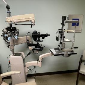
- Slit‑lamp exam: carefully inspect lash bases for cylindrical collarettes; evaluate lid margin for telangiectasia, MGD signs; assess conjunctiva and cornea with fluorescein.
- The presence of collarettes in symptomatic blepharitis strongly supports Demodex involvement [1][2].
Imaging and laboratory testing
- Lash epilation and light microscopy can demonstrate mites and eggs; useful in atypical or refractory cases but not routinely required [1].
- In vivo confocal microscopy visualizes mites within follicles/glands; mainly research.
- Bacterial cultures only if secondary infection suspected; not part of standard workup.
Differential diagnosis
| Entity | Distinguishing features |
|---|---|
| Staphylococcal blepharitis | Crusts and flakes along lashes without cylindrical sleeves; ulcerative collarettes with frank ulceration; responds to topical antibiotic ointments. |
| Seborrheic blepharitis | Greasy scales along lashes and brows; seborrhea of scalp; minimal lid margin ulceration. |
| Phthiriasis palpebrarum (lice) | Visible nits/crab lice at lash bases; intense pruritus; fluoresce on Wood lamp; sexual or close contact history. |
| Allergic blepharoconjunctivitis | Itching predominant; papillary conjunctivitis; seasonal or atopic history; lacks Demodex collarettes. |
| Ocular rosacea without Demodex predominance | Lid telangiectasia, MGD, facial flushing; may coexist with Demodex. |
| Herpetic blepharitis | Vesicles/ulcers on lid skin; dendritic keratitis if cornea involved; viral prodrome. |
| Bacterial conjunctivitis | Mucopurulent discharge, conjunctival papillae; minimal lid margin findings early; sudden onset. |
| Atopic dermatitis involving lids | Lichenified, eczematous lids; atopic diathesis; Dennie‑Morgan folds. |
Treatment
Medical management
- Lotilaner ophthalmic solution 0.25% (Xdemvy): FDA‑approved for Demodex blepharitis. Dose 1 drop in each eye bid (approximately every 12 hours) for 6 weeks [4]. Advise contact lens removal before instillation and reinsert after the interval specified in the label [4]. Common adverse effects: instillation site stinging/burning, redness.
- Lid hygiene (adjunct): warm compresses 5–10 minutes followed by gentle lid margin cleansing to remove collarettes and express meibum; preservative‑free artificial tears for surface symptoms.
- Topical anti‑inflammatory therapy (adjunct, off‑label): short course of low‑potency corticosteroid (eg, loteprednol ophthalmic) for significant inflammation or keratitis; avoid prolonged use; consider cyclosporine or lifitegrast for chronic ocular surface inflammation when indicated [2].
- Management of comorbid ocular rosacea/MGD (off‑label where noted): doxycycline 50–100 mg PO bid for 2–4 weeks, then 50 mg daily for 4–8 weeks as needed; contraindicated in pregnancy/young children; subantimicrobial dosing options exist [2].
- Tea tree oil (terpinen‑4‑ol)–based lid cleansers (off‑label): may reduce mite load; avoid high‑concentration products in the eye due to epithelial toxicity; evidence quality is variable and irritation is common [1][2].
- Topical ivermectin 1% cream to lash line (off‑label): applied carefully to lid margins (typically qhs) for several weeks may reduce mites; avoid ocular instillation; local irritation possible [1].
Procedural and surgical management
- Microblepharoexfoliation (in‑office mechanical debridement): removes collarettes and biofilm from the lid margin; used as an adjunct to medical therapy in refractory cases [2].
- Meibomian gland expression and thermal pulsation: adjuncts to address MGD in D. brevis–predominant disease [2].
- Incision and curettage for persistent chalazia; address underlying Demodex load to reduce recurrence.
Special populations
- Pediatric: Demodex blepharitis occurs in children and may present with recurrent chalazia or blepharokeratoconjunctivitis. Lotilaner ophthalmic solution is approved for pediatric patients aged ≥2 years [4]. Avoid doxycycline; use gentle lid hygiene; exercise caution with tea tree oil products due to irritation risk.
- Pregnancy/lactation: Safety data for lotilaner in pregnancy/lactation are limited; consider supportive lid hygiene and defer nonessential therapies; avoid systemic tetracyclines [4][2]. Shared decision‑making is essential.
- Immunocompromised: Higher mite burdens may occur; coordinate with dermatology for concomitant facial demodicosis; monitor closely for secondary infections.
Prognosis
- With targeted therapy and ongoing lid hygiene, symptoms and collarettes typically improve within 6–8 weeks (covering mite life cycles). Relapses can occur; maintenance lid care reduces recurrence [1][2][4].
- Prognosis is guarded in longstanding MGD with gland dropout; ocular surface disease may persist and require chronic management.
Prevention and Patient Counseling
- Daily lid hygiene (warm compresses and gentle margin cleansing) helps prevent recurrence.
- Replace eye makeup regularly; avoid sharing cosmetics or towels; wash pillowcases and bedding in hot water.
- Manage facial rosacea and seborrheic dermatitis in collaboration with dermatology.
- Contact lens wearers should optimize lens hygiene and consider reducing wear during active treatment.
When to Seek Immediate Care
- Sudden decrease in vision or severe eye pain.
- Marked light sensitivity, new corneal haze, or a white spot on the cornea.
- Rapidly worsening redness with swelling of the eyelids or surrounding skin.
- Fever, spreading skin infection, or inability to open the eye.
- Symptoms after eye surgery or trauma.
References
- AAO EyeWiki. Demodex Blepharitis. https://eyewiki.aao.org/Demodex_Blepharitis. Accessed July 24, 2026.
- American Academy of Ophthalmology. Blepharitis Preferred Practice Pattern. 2023. https://www.aao.org/preferred-practice-pattern/blepharitis-ppp. Accessed July 24, 2026.
- National Eye Institute. Blepharitis. https://www.nei.nih.gov/learn-about-eye-health/eye-conditions-and-diseases/blepharitis. Accessed July 24, 2026.
- DailyMed. XDEMVY (lotilaner) ophthalmic solution 0.25%—prescribing information. https://dailymed.nlm.nih.gov. Accessed July 24, 2026.
- StatPearls. Blepharitis. Treasure Island (FL): StatPearls Publishing; 2026. https://www.ncbi.nlm.nih.gov/books/NBK459305/. Accessed July 24, 2026.
- Kheirkhah A, Casas V, Li W, Raju VK, Tseng SC. Ocular demodicosis: Clinical implications and treatment. Ocul Surf. 2020;18(2):363-377. doi:
ICD‑10‑CM
- B88.0 Demodicosis (other acariasis)
- H01.00–H01.05 Blepharitis subcodes with eyelid and laterality specification (eg, H01.001 right upper eyelid, unspecified blepharitis; H01.004 left upper eyelid, unspecified blepharitis). Use the specific code that matches type and eyelid involved per ICD‑10‑CM tabular list.
CPT (when applicable)
- 67800 Excision of chalazion, single.
- 65205 Removal of foreign body, external eye; conjunctival superficial (if indicated for corneal/ conjunctival involvement).
- 65778 Placement of amniotic membrane (only if used for severe keratitis; uncommon in Demodex).
Disclaimer: This article is for informational purposes and is not a substitute for professional medical advice, diagnosis, or treatment. Always consult a qualified eye care professional about your specific condition. If you have sudden vision loss, severe eye pain, or an eye injury, seek emergency care immediately.