Farsightedness (hyperopia) is a refractive error in which parallel light focuses behind, rather than on, the retina when accommodation (focusing effort) is relaxed. It typically causes near blur, eye strain, and headaches with sustained near work; higher degrees can blur distance vision. Diagnosis is by refraction, often with cycloplegia (temporarily relaxing accommodation), and ocular alignment assessment in children. Treatment is optical (spectacles or contact lenses); laser or lens-based surgery may be options for selected adults.

Key Points

  • Farsightedness (hyperopia) results from a short axial length or insufficient optical power of the cornea/lens; the eye must accommodate to see clearly, especially at near.
  • Children can mask hyperopia via strong accommodation, risking asthenopia (eye strain), amblyopia, and accommodative esotropia if uncorrected.
  • Cycloplegic refraction is required to uncover latent hyperopia in pediatric patients and when symptoms exceed the measured error.
  • Hyperopia is a risk factor for primary angle-closure disease due to shallow anterior chambers in some eyes.
  • Management is primarily plus-lens correction; full correction is indicated in accommodative esotropia, while partial correction may be used in asymptomatic children without strabismus.
  • Hyperopic LASIK/PRK can help low to moderate hyperopia but has higher regression and predictability challenges than myopic treatments.

Anatomy and Physiology

The eye’s refractive power is determined mainly by the cornea and crystalline lens. The retina must receive a focused image at the fovea for clear vision. In emmetropia, parallel rays focus on the retina at rest. Accommodation, mediated by ciliary muscle contraction and lens shape change, adds optical power for near tasks. Anterior chamber depth and axial length influence refractive state; shorter axial length or flatter cornea reduces total power.

Etiology

  • Axial hyperopia: Axial length shorter than average; the most common cause.
  • Refractive (index/curvature) hyperopia:
    • Flat corneal curvature.
    • Reduced lens power (eg, postsurgical, aphakia).
    • Lenticular changes (eg, early lens sclerosis usually induces myopia, but post-cataract extraction causes hyperopia without IOL).
  • Functional/physiologic: Newborns are commonly hyperopic; emmetropization reduces hyperopia over early childhood.
  • Secondary:
    • After corneal refractive surgery for myopia if overcorrected.
    • Ocular inflammation or medications rarely altering accommodation.

Genetic and familial factors contribute to refractive development.

Pathophysiology

In hyperopia, the eye’s resting focus lies behind the retinal plane. Clear vision requires accommodation even at distance for higher hyperopia and at near for mild to moderate hyperopia. Sustained accommodation drives accommodative convergence and can precipitate esodeviation (accommodative esotropia) in susceptible children. Latent hyperopia is masked by tonic ciliary muscle activity and becomes manifest with cycloplegia or fatigue. With aging, presbyopia reduces accommodative reserve, unmasking previously asymptomatic hyperopia.

Hyperopic eyes may have shallower anterior chambers and narrower angles, increasing susceptibility to pupillary block and angle-closure mechanisms.

Epidemiology

Hyperopia is common in the United States across the lifespan. Mild hyperopia is frequent in infancy and early childhood and declines with emmetropization through school age; a portion persists into adulthood. Clinically significant hyperopia occurs in both children and adults and varies by age and ancestry [1,2,3]. Precise prevalence estimates vary by study and threshold.

Classification

  • By magnitude (spherical equivalent):
    • Low: ≤ +2.00 D
    • Moderate: +2.25 to +5.00 D
    • High: > +5.00 D
  • By mechanism: axial vs refractive (corneal curvature or lenticular).
  • By accommodative status:
    • Latent hyperopia: masked without cycloplegia.
    • Manifest hyperopia: evident on routine refraction.
    • Facultative: can be overcome by accommodation.
    • Absolute: cannot be overcome; persistent blur without optical correction.
  • By symmetry: isometropic (both eyes similar) vs anisometropic (asymmetric).

Symptoms and Signs of Farsightedness

  • Adults and adolescents

    • Near blur and fluctuating vision, worse after prolonged near work.
    • Asthenopia: frontal/ocular headaches, brow ache, eye fatigue.
    • Intermittent distance blur, especially when tired or with uncorrected moderate/high hyperopia.
    • Reduced accommodation amplitudes with age (presbyopia) exacerbate symptoms.
  • Children

    • May be asymptomatic due to robust accommodation.
    • Behavioral signs: avoidance of near tasks, short attention with reading, rubbing eyes.
    • Strabismus: intermittent or constant esotropia with near fixation (accommodative esotropia).
    • Amblyopia: reduced best-corrected acuity in one or both eyes with high or asymmetric hyperopia.
  • Examination findings

    • Reduced unaided near acuity; distance acuity normal or subnormal depending on magnitude and accommodative reserve.
    • Esophoria or esotropia at near; high accommodative convergence/accommodation (AC/A) ratio in some.
    • Narrow anterior chamber angles on gonioscopy in some hyperopic eyes.

Complications

  • Amblyopia (isoametropic or anisometropic) if significant hyperopia is uncorrected in early childhood [2].
  • Accommodative esotropia and associated binocular vision anomalies.
  • Asthenopia impacting school/work performance.
  • Angle-closure suspect status or angle-closure glaucoma risk in anatomically predisposed eyes [4].
  • Contact lens intolerance or dry eye symptoms with over-accommodation; fluctuating blur overlapping with dry eye disease.

Diagnosis

Clinical evaluation

  • History: visual blur pattern (near vs distance), asthenopia, reading endurance, diplopia/eye turn in children, family history.
  • Visual acuity: distance and near, with pinhole.
  • Refraction:
    • Objective retinoscopy and/or autorefraction.
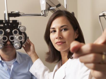
    • Cycloplegic refraction to reveal latent hyperopia, essential in children and whenever symptoms exceed manifest findings (eg, cyclopentolate 1% 1 drop in each eye, possibly repeated in 5 minutes; check refraction after 30–45 minutes) [1,5].
  • Ocular alignment and binocularity:
    • Cover–uncover and alternate cover tests at distance and near; evaluate AC/A ratio when indicated.
    • Stereopsis testing in children.
  • Anterior segment/anatomy:
    • Slit-lamp exam; estimate anterior chamber depth.
    • Gonioscopy in adults with symptoms of halos, shallow chambers, or narrow angles on exam.
  • Fundus exam to exclude amblyopic sequelae or other pathology.

Imaging and laboratory testing

  • Not routinely required.
  • Anterior segment optical coherence tomography (AS-OCT) or ultrasound biomicroscopy can quantify angle anatomy in suspected angle narrowing.
  • Axial length biometry can document mechanism (axial vs refractive) when clinically relevant (eg, preoperative refractive surgery planning).

No laboratory tests are indicated.

Differential diagnosis

Entity Distinguishing features
Presbyopia Age >40–45; normal distance refraction; near blur relieved by near add; accommodative amplitude reduced for age.
Astigmatism Blur at all distances; improves with cylindrical correction; keratometry/topography abnormal.
Accommodative insufficiency Near blur/asthenopia in teens/young adults with normal refraction; low accommodative amplitude; near add relieves symptoms.
Convergence insufficiency Near diplopia/eye strain; normal refraction; receded near point of convergence; improved with vergence therapy.
Dry eye disease Fluctuating vision, burning, worse with screens; improves with blinking or lubricants; corneal staining present. See dry eye content.
Accommodative spasm (pseudomyopia) Variable refraction, miosis, excess convergence; minus shift on refraction; relieved by cycloplegia.
Keratitis Pain, photophobia, focal corneal opacity; urgent evaluation required.
Ocular migraine (visual aura) Transient scintillating scotoma or zig-zag lines; normal ocular exam between episodes.

Related conditions on this site:

  • Dry eye symptoms can mimic intermittent blur; see our overview of dry eye syndrome (/blog/dry-eye-syndrome).
  • Painful red eye with photophobia suggests keratitis, not refractive error (/disease/keratitis).
  • Eyelid lumps like chalazion can cause localized irritation but do not cause refractive blur (/disease/chalazion).
  • Mucopurulent discharge and diffuse redness suggest bacterial conjunctivitis, not farsightedness (/disease/bacterial-conjunctivitis).

Treatment

Medical management

  • Optical correction (first-line)
    • Spectacles: Plus spherical lenses (single-vision for distance or near; bifocal/progressive add for presbyopic adults). In accommodative esotropia, prescribe full cycloplegic hyperopic correction to align the eyes; bifocal add for high AC/A ratio may reduce near esodeviation [2].
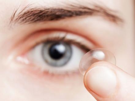
    • Contact lenses: Soft or rigid gas-permeable options provide full-time correction; consider for anisometropia or high hyperopia to reduce image size differences (aniseikonia).
  • Diagnostic/adjunct pharmacology
    • Cycloplegics for refraction (eg, cyclopentolate 1% as above; 0.5% in infants) [1,5].
    • Amblyopia therapy when indicated (eg, patching or atropine 1% penalization, per Pediatric Eye Disease Investigator Group protocols) after appropriate refractive correction [2]. Amblyopia therapy addresses a complication, not hyperopia per se.

Notes:

  • In asymptomatic children without strabismus, some clinicians partially correct moderate hyperopia to balance clarity and emmetropization potential; practice varies and should follow pediatric guidelines [2]. Exact diopter thresholds depend on age, symmetry, and symptoms.
  • Monitor for accommodative symptoms and school performance; adjust prescriptions accordingly.

Procedural and surgical management

  • Corneal laser vision correction (adults)
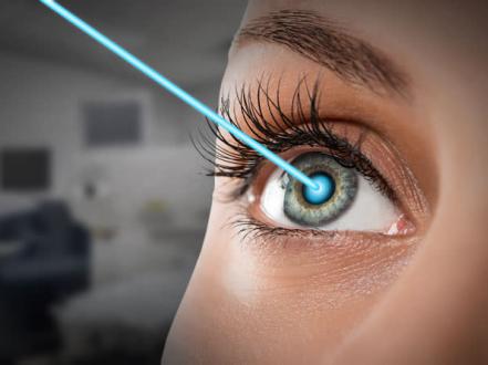
    • LASIK (laser in situ keratomileusis) and PRK (photorefractive keratectomy) are FDA-approved for low to moderate hyperopia in selected ranges depending on platform (commonly up to about +4.00 to +6.00 D with or without astigmatism) [6]. Outcomes for hyperopia have greater variability and regression risk than for myopia; careful patient selection and counseling are essential [1,6].
    • SMILE (small-incision lenticule extraction) is not FDA-approved for hyperopia as of July 2026 [6].
  • Lens-based options
    • Refractive lens exchange (clear lens extraction with intraocular lens) may be considered in high hyperopia, especially in presbyopic adults or those with narrow angles where lens extraction can deepen the angle [4]. Discuss risks (retinal detachment risk is lower than in high myopia but includes endophthalmitis, dysphotopsias).
    • Phakic IOLs for hyperopia have limited availability/indications in the US; lens-based correction should be considered case by case [1,6].
  • Obsolescent/limited
    • Conductive keratoplasty for low hyperopia has largely fallen out of favor due to regression and instability [1].

Procedures are elective and typically not covered by insurance. Preoperative evaluation includes stable refraction, corneal tomography, tear film assessment, and pupil/angle evaluation.

Special populations

  • Pediatric
    • Perform cycloplegic refraction in all children evaluated for suspected hyperopia, strabismus, or amblyopia.
    • Prescribe full hyperopic correction for accommodative esotropia; consider bifocals for high AC/A ratio [2].
    • In otherwise asymptomatic children without strabismus, prescribing thresholds vary by age and magnitude; follow AAPOS-informed guidance and monitor for amblyopia risk [2,3].
  • Pregnancy/lactation
    • Corneal thickness, tear film, and refraction can fluctuate; defer new refractive surgery until several months postpartum; update spectacle/contact prescriptions if needed for function [6].
  • Older adults
    • Presbyopia unmasking increases need for plus correction at near and sometimes at distance; evaluate for narrow angles and counsel regarding angle-closure symptoms [4].

Prognosis

With appropriate optical correction, visual function and comfort are excellent. In children, timely correction reduces the risk of amblyopia and can control accommodative esotropia. Hyperopic laser procedures can reduce dependence on glasses for selected adults but have higher regression and enhancement rates than myopic treatments [1,6]. Hyperopia-associated narrow angles warrant surveillance to mitigate angle-closure risk [4].

Prevention and Patient Counseling

  • Hyperopia cannot be prevented; it reflects ocular anatomy and development. Early detection via vision screening and comprehensive eye exams enables timely correction and amblyopia prevention [2,3].
  • Counsel on:
    • Proper spectacle fit and full-time wear when prescribed for strabismus/amblyopia risk.
    • Contact lens hygiene to avoid infectious keratitis.
    • Near-work ergonomics and regular breaks to manage asthenopia.
    • Recognizing symptoms of angle closure (halos, severe eye pain, headache, nausea) and seeking urgent care.

When to Seek Immediate Care

  • Severe eye pain, headache, halos around lights, and nausea/vomiting (possible angle-closure attack).
  • Sudden vision loss or new double vision.
  • A red, painful eye with light sensitivity or discharge.
  • A new eye turn (crossing) in a child or loss of alignment with glasses.
  • Eye injury or acute contact lens–related pain.

References

  1. American Academy of Ophthalmology. Refractive Errors & Refractive Surgery Preferred Practice Pattern. San Francisco, CA: AAO; 2022. https://www.aao.org/preferred-practice-pattern/refractive-errors-ppp

  2. American Association for Pediatric Ophthalmology and Strabismus (AAPOS). Refractive Errors in Children. 2024. https://aapos.org/glossary/refractive-errors-in-children

  3. US Preventive Services Task Force. Vision Screening in Children Aged 3 to 5 Years: US Preventive Services Task Force Recommendation Statement. JAMA. 2017;318(9):836-844. doi:10.1001/jama.2017.11260

  4. EyeWiki (AAO). Primary Angle Closure Glaucoma. 2024. https://eyewiki.org/Primary_Angle_Closure_Glaucoma

  5. StatPearls. Cycloplegics. Treasure Island (FL): StatPearls Publishing; 2024. https://www.ncbi.nlm.nih.gov/books/NBK555932/

  6. StatPearls. Laser In Situ Keratomileusis (LASIK). Treasure Island (FL): StatPearls Publishing; 2025. https://www.ncbi.nlm.nih.gov/books/NBK538291/

  7. National Eye Institute (NIH). Facts About Refractive Errors. 2024. https://www.nei.nih.gov/learn-about-eye-health/eye-conditions-and-diseases/refractive-errors

  8. EyeWiki (AAO). Hyperopia. 2024. https://eyewiki.org/Hyperopia

ICD-10-CM

  • H52.01 Hypermetropia, right eye
  • H52.02 Hypermetropia, left eye
  • H52.03 Hypermetropia, bilateral
  • H52.00 Hypermetropia, unspecified eye

Relevant procedures (professional tier)

  • 92015 Refraction
  • 92060 Sensorimotor exam
  • 92020 Gonioscopy

Byline Author: [AUTHOR NAME], [CREDENTIALS PENDING] Medical reviewer: [REVIEWER NAME], [CREDENTIALS PENDING] Reviewed: July 2026 Last updated: July 2026

Disclaimer This article is for informational purposes and is not a substitute for professional medical advice, diagnosis, or treatment. Always consult a qualified eye care professional about your specific condition. If you have sudden vision loss, severe eye pain, or an eye injury, seek emergency care immediately.