Phlegmon is a diffuse, spreading, suppurative inflammation of soft tissue without a well-formed abscess capsule. In eye care, phlegmon most often involves the eyelids (preseptal) or the orbit (postseptal) as part of the orbital cellulitis spectrum. Patients present with eyelid edema, erythema, and tenderness; orbital involvement causes proptosis, pain with eye movements, ophthalmoplegia, and sometimes decreased vision. Diagnosis is clinical, supported by contrast-enhanced CT or MRI when orbital disease or complications are suspected. Treatment is prompt systemic antibiotics; surgical drainage is indicated if a discrete abscess forms or vision is threatened.

Key Points

  • Phlegmon denotes diffuse purulent cellulitis without a capsule; in the orbit, it precedes or accompanies subperiosteal/orbital abscess.
  • Sinusitis, especially ethmoiditis, is the leading source of orbital phlegmon in children; trauma, skin infection, and hordeolum/stye are common sources of preseptal disease.
  • Orbital involvement is an emergency; red flags are proptosis, painful/restricted eye movements, decreased vision, afferent pupillary defect, fever, and severe headache.
  • Contrast-enhanced CT of the orbits/sinuses distinguishes phlegmon (ill-defined enhancement, fat stranding) from abscess (rim-enhancing collection) and identifies complications.
  • Management: preseptal phlegmon usually responds to oral antibiotics; orbital phlegmon requires hospital admission, IV broad-spectrum antibiotics, ENT/ophthalmology co-management, and surgical drainage if an abscess develops or vision is compromised.
  • Fungal orbital phlegmon (e.g., mucormycosis) must be considered in immunocompromised or diabetic patients and demands urgent antifungal therapy and debridement.

Anatomy and Physiology

The orbital septum is a fibrous barrier extending from the orbital rim to the eyelid margin, separating preseptal tissues (skin, orbicularis, subcutaneous tissue) from postseptal orbital contents (extraocular muscles, fat, optic nerve, globe). Paranasal sinuses, particularly the ethmoids, share thin bony partitions with the orbit and communicate via valveless veins, allowing rapid spread of infection into the orbit and cavernous sinus.

Etiology

  • Bacterial sources
    • Paranasal sinusitis (ethmoid > maxillary/frontal): streptococci, anaerobes; in some series, Staphylococcus aureus (including MRSA), Streptococcus pneumoniae, group A streptococci, and mixed anaerobes [1,2].
    • Eyelid/skin infections: hordeolum/stye, impetigo, trauma, insect bites.
    • Postsurgical or posttraumatic inoculation.
    • Dacryocystitis or dacryoadenitis.
  • Less common
    • Fungal infections: mucormycosis, aspergillosis (diabetes, neutropenia).
    • Odontogenic infections (maxillary molars).
  • Vaccination has markedly reduced Haemophilus influenzae type b–associated cases in children [3].

Pathophysiology

Phlegmon represents acute, suppurative cellulitis with edema, leukocyte infiltration, and tissue necrosis without a mature capsule. In the orbit, infection spreads from the ethmoid sinuses through dehiscent lamina papyracea or via venous channels. Inflammation increases intraorbital pressure, impairs venous outflow, and can compromise the optic nerve and retinal perfusion. Progression may produce a subperiosteal or intraconal abscess; further spread risks cavernous sinus thrombosis and intracranial complications.

Epidemiology

  • Preseptal cellulitis is more common than orbital cellulitis across all ages; orbital phlegmon peaks in children <15 years, often secondary to sinusitis [1,2].
  • After Hib vaccination, H. influenzae accounts for a small minority of pediatric cases in the US [3].
  • Community-acquired MRSA prevalence varies geographically; empiric coverage is guided by local patterns [4].

Classification

Phlegmon within the orbit is commonly described using the Chandler classification of sinusitis-related orbital complications [5]:

  • Group 1: Preseptal cellulitis (preseptal phlegmon)
  • Group 2: Orbital cellulitis (orbital phlegmon without abscess)
  • Group 3: Subperiosteal abscess
  • Group 4: Orbital abscess
  • Group 5: Cavernous sinus thrombosis

Radiologically, phlegmon shows ill-defined, contrast-enhancing inflamed tissue and fat stranding, while abscess appears as a low-attenuation collection with rim enhancement and diffusion restriction on MRI [2].

Symptoms and Signs

  • Preseptal phlegmon (preseptal cellulitis)

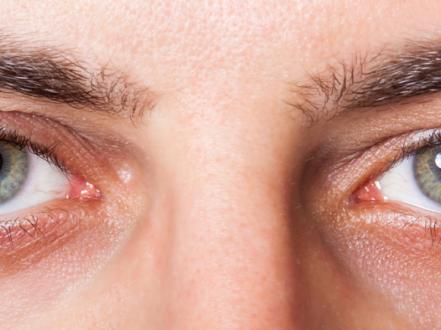
    • Eyelid erythema, warmth, tenderness, and edema.
    • Normal visual acuity, no pain with eye movements, no ophthalmoplegia, no proptosis.
    • Often history of hordeolum, skin lesion, or trauma.
  • Orbital phlegmon (postseptal/orbital cellulitis)

    • Fever, malaise, orbital pain.
    • Proptosis, chemosis, painful and/or limited extraocular movements.
    • Decreased vision or color desaturation; relative afferent pupillary defect (optic nerve compromise).
    • Headache, sinus tenderness; in severe cases, systemic toxicity or neurologic symptoms.
  • Fungal orbital phlegmon (high-risk hosts)

    • Rapid progression, necrotic eschar of nasal turbinates or palate (mucormycosis), cranial neuropathies.

Complications

  • Subperiosteal or orbital abscess (vision-threatening).
  • Optic neuropathy, central retinal artery occlusion.
  • Cavernous sinus thrombosis, meningitis, epidural/subdural empyema, brain abscess.
  • Lacrimal drainage obstruction or chronic sinus disease.
  • Sepsis (rare with prompt care).

Diagnosis

Clinical evaluation

  • History: onset relative to sinusitis, trauma, skin lesions, dental disease, or surgery; systemic symptoms; immunocompromise/diabetes.
  • Examination:
    • Distinguish preseptal from orbital involvement: assess vision, pupils, ocular motility, presence of proptosis and chemosis.
    • Evaluate for meningeal signs and cranial neuropathies in severe cases.

Imaging and laboratory testing

  • Imaging:
    • Contrast-enhanced CT orbits and paranasal sinuses is first-line when orbital phlegmon is suspected, when exam is limited (young child), or if no improvement within 24–48 hours of antibiotics [1,2].
    • MRI orbits/brain with contrast is preferred for suspected intracranial spread, cavernous sinus thrombosis, fungal disease, or when iodinated contrast or radiation is contraindicated.
  • Laboratory:
    • CBC and inflammatory markers (CRP/ESR) to assess severity and response.
    • Blood cultures in febrile or toxic patients; wound/abscess cultures if drainage is present or obtained.
    • Nasal/sinus cultures are not routinely diagnostic for orbital disease.

Differential diagnosis

Entity Distinguishing features
Chalazion/hordeolum Localized eyelid nodule; focal tenderness; no fever, no orbital signs; may point to lid margin; see chalazion: https://myvisioncare.org/disease/chalazion
Allergic angioedema Bilateral, non-tender, pruritic edema; history of allergen exposure; normal ocular motility/vision
Bacterial conjunctivitis Conjunctival hyperemia and purulent discharge; minimal lid tenderness; preserved motility/vision; see bacterial conjunctivitis: https://myvisioncare.org/disease/bacterial-conjunctivitis
Keratitis Photophobia, foreign body sensation, corneal opacity/ulcer; pain without marked lid erythema; risk with contact lenses; see keratitis: https://myvisioncare.org/disease/keratitis
Sinusitis without orbital involvement Facial pain/pressure, rhinorrhea; normal ocular exam
Carotid–cavernous fistula Pulsatile proptosis, bruit, corkscrew conjunctival vessels, chronic course
Thyroid eye disease Painless proptosis, lid retraction, restrictive myopathy; euthyroid/hyperthyroid history
Herpes zoster ophthalmicus Dermatomal vesicular rash (V1), neuropathic pain; may secondarily infect

Treatment

Medical management

  • Preseptal phlegmon (outpatient, if mild and reliable follow-up)

    • Empiric oral antibiotics targeting streptococci and staphylococci; include MRSA coverage where prevalent or if risk factors are present [4].
    • Amoxicillin-clavulanate (Augmentin) 875/125 mg PO bid for 5–7 days (children: 45 mg/kg/day amoxicillin component PO divided bid).
    • If MRSA risk or beta-lactam allergy: trimethoprim-sulfamethoxazole (Bactrim DS) 1–2 tablets PO bid (children: 8–12 mg/kg/day TMP divided bid) plus amoxicillin or clindamycin 300 mg PO q6–8h (children: 10–13 mg/kg PO q8h). Doxycycline 100 mg PO bid is an alternative in adults; avoid in children <8 years and pregnancy.
    • Warm compresses; treat underlying hordeolum/skin source; close recheck in 24–48 hours.
    • Escalate to imaging and IV therapy if no improvement or if orbital signs emerge.
  • Orbital phlegmon (inpatient)

    • Urgent ophthalmology and otolaryngology consultation.
    • IV broad-spectrum antibiotics covering streptococci, staphylococci (including MRSA if indicated), gram-negatives, and anaerobes pending cultures [1,2,4]:
    • Ampicillin-sulbactam 3 g IV q6h or piperacillin-tazobactam 4.5 g IV q6–8h.
    • Plus vancomycin (dose per pharmacy/Nomogram; typically 15–20 mg/kg IV q8–12h) if MRSA risk or severe infection.
    • Alternative cephalosporin-based regimens: ceftriaxone 2 g IV q24h or cefotaxime 2 g IV q8h plus metronidazole 500 mg IV q8h for anaerobic coverage.
    • Penicillin allergy (severe): levofloxacin 750 mg IV q24h plus metronidazole 500 mg IV q8h; add vancomycin if MRSA risk.
    • Pediatrics (examples; adjust per weight and renal function): ampicillin-sulbactam 200–300 mg/kg/day IV divided q6h; ceftriaxone 50 mg/kg/day IV; vancomycin 15 mg/kg IV q6h; metronidazole 30 mg/kg/day IV divided q8h.
    • Manage pain, nasal decongestion/saline irrigations for sinusitis (ENT-directed). Systemic corticosteroids are not first-line and, if used, should be started only after antibiotics and with subspecialty guidance [2].
    • Monitor vision, pupils, intraocular pressure, and motility at least daily.

Procedural and surgical management

  • Indications for drainage (ophthalmology/ENT co-management) [1,2,5,6]:
    • Declining vision or afferent pupillary defect at any time.
    • Large or expanding subperiosteal/orbital abscess on imaging.
    • Frontal sinusitis, gas in collection, or intracranial complications.
    • No clinical improvement or worsening after 24–48 hours of appropriate IV antibiotics.
    • Suspicion for anaerobic/odontogenic source or foreign body.
  • Approaches:
    • Endoscopic sinus surgery with subperiosteal abscess drainage (ENT).
    • Orbitotomy with abscess drainage when intraconal collections or lateral/posterior abscesses are not endoscopically accessible (CPT 67400).
    • Obtain aerobic/anaerobic/fungal cultures during drainage to tailor therapy.
  • Fungal disease
    • Urgent surgical debridement plus high-dose systemic antifungals (e.g., liposomal amphotericin B) in consultation with infectious disease and ENT/neurosurgery [2].

Special populations

  • Pediatric
    • Higher incidence; Hib rare post-vaccine. Lower threshold for imaging when exam is limited. Weight-based antibiotic dosing; avoid doxycycline in children <8 years.
  • Pregnancy/lactation
    • Avoid tetracyclines and fluoroquinolones; beta-lactams preferred. Coordinate with obstetrics.
  • Immunocompromised/diabetes
    • Broader differential including mucormycosis; early MRI and ENT evaluation; aggressive surgical and antifungal management when suspected.
  • Elderly
    • Consider atypical presentations and polypharmacy; adjust antibiotic dosing for renal function.

Prognosis

With timely diagnosis and appropriate antibiotics, most preseptal and orbital phlegmon resolves without sequelae. Delayed treatment, resistant organisms, intracranial spread, fungal infection, or compressive optic neuropathy worsen outcomes and increase the risk of permanent vision loss or neurologic complications. Recurrence is uncommon if the sinus or skin source is effectively treated.

Prevention and Patient Counseling

  • Prompt evaluation of eyelid infections and bacterial sinusitis.
  • Manage hordeola and blepharitis to reduce eyelid infection risk; consider warm compress hygiene and lid care.
  • Maintain up-to-date Hib and pneumococcal vaccination in children [3].
  • Protect periocular skin from trauma; treat dental infections.
  • For contact lens users with red, painful eyes, remove lenses and seek eye care to exclude keratitis rather than self-treat; see our overview on dry eye for noninfectious irritation: https://myvisioncare.org/blog/dry-eye-syndrome

When to Seek Immediate Care

  • Fever with a swollen, red, and painful eyelid, especially in a child.
  • Eye bulging, double vision, or pain with moving the eye.
  • Sudden change in vision, color desaturation, or unequal pupils.
  • Severe headache, vomiting, confusion, or stiff neck.
  • Black scab in the nose or palate in a person with diabetes or weakened immunity.
  • No improvement within 24–48 hours of starting antibiotics.

References

  1. Chaudhry IA, Al-Rashed W, Arat YO. The Hot Orbit: Orbital Cellulitis. Middle East Afr J Ophthalmol. 2012;19(1):34-42. doi:10.4103/0974-9233.92114.
  2. Nageswaran S, Woods CR, Benjamin DK Jr, Givner LB, Shetty AK. Orbital cellulitis in children. Pediatr Infect Dis J. 2006;25(8):695-699. doi:10.1097/01.inf.0000226938.13299.43.
  3. CDC. Haemophilus influenzae Type b (Hib) Vaccination: What Everyone Should Know. Updated 2023. https://www.cdc.gov/vaccines/vpd/hib/public/index.html
  4. Stevens DL, Bisno AL, Chambers HF, et al. Practice Guidelines for the Diagnosis and Management of Skin and Soft Tissue Infections: 2014 Update by the IDSA. Clin Infect Dis. 2014;59(2):e10-e52. doi:10.1093/cid/ciu444.
  5. Chandler JR, Langenbrunner DJ, Stevens ER. The pathogenesis of orbital complications in acute sinusitis. Laryngoscope. 1970;80(9):1414-1428. doi:10.1288/00005537-197009000-00007.
  6. Rudloe TF, Harper MB, Prabhu SP, Rahbar R, Vanderveen D, Kimia AA. Acute Periorbital Infections: Who Needs Emergent Imaging? Pediatrics. 2010;125(4):e719-e726. doi:10.1542/peds.2009-1084.
  7. AAO EyeWiki. Orbital Cellulitis. American Academy of Ophthalmology. https://eyewiki.aao.org/Orbital_Cellulitis
  8. StatPearls. Periorbital Cellulitis. Updated 2024. https://www.ncbi.nlm.nih.gov/books/NBK470408/
  9. StatPearls. Orbital Cellulitis. Updated 2024. https://www.ncbi.nlm.nih.gov/books/NBK507901/
  10. Chow AW, Benninger MS, Brook I, et al. IDSA Clinical Practice Guideline for Acute Bacterial Rhinosinusitis in Children and Adults. Clin Infect Dis. 2012;54(8):e72-e112. doi:10.1093/cid/cis370.

Disclaimer: This article is for informational purposes and is not a substitute for professional medical advice, diagnosis, or treatment. Always consult a qualified eye care professional about your specific condition. If you have sudden vision loss, severe eye pain, or an eye injury, seek emergency care immediately.