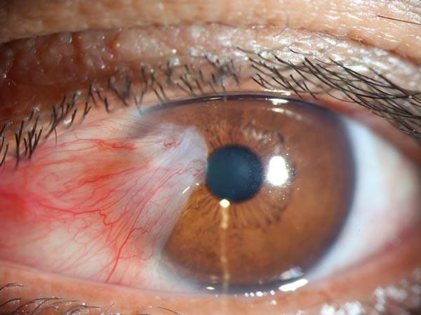
A pterygium is a benign, wing-shaped fibrovascular growth of bulbar conjunctiva that extends across the limbus onto the corneal surface. It is driven by chronic ultraviolet (UV) exposure and ocular surface irritation. Typical symptoms are cosmetic redness, dryness, foreign-body sensation, and, when large, decreased vision from induced astigmatism or encroachment on the visual axis. Diagnosis is clinical at the slit lamp. Initial treatment is UV protection and lubricants; surgery is indicated for vision threat, significant astigmatism, recurrent inflammation, restrictive diplopia, dellen, or unacceptable cosmesis.

Key Points

  • Pterygium is a UV-related conjunctival proliferation that migrates onto the cornea; progression is usually slow but may be recurrent after excision.
  • Symptoms include irritation, redness, and dryness; vision is affected by irregular astigmatism or direct involvement of the visual axis.
  • Slit-lamp examination is diagnostic; corneal topography quantifies astigmatism, and anterior segment optical coherence tomography (AS-OCT) can document depth and extent.
  • First-line management is environmental protection (UV-blocking eyewear, hats) and ocular surface optimization (artificial tears; short steroid courses for flares).
  • Excision with conjunctival autografting (often with fibrin glue) is the standard surgical approach; judicious mitomycin C use lowers recurrence but carries risk.
  • Distinguish from pinguecula (no corneal extension), pseudopterygium (postinflammatory adhesion), and ocular surface squamous neoplasia (OSSN).

Anatomy and Physiology

The conjunctiva is a non-keratinized mucous membrane covering the sclera (bulbar conjunctiva) and inner eyelids (palpebral conjunctiva). The limbus houses corneal epithelial stem cells. Chronic UV-B exposure induces elastotic degeneration of the bulbar conjunctival stroma (solar elastosis) and alters limbal stem cell homeostasis. The cornea is avascular; conjunctival fibrovascular tissue crossing the limbus disrupts corneal optics and tear film stability.

Etiology

  • Chronic UV-A/UV-B exposure (outdoor work, tropical/subtropical latitudes)
  • Environmental irritants (wind, dust, dry climates)
  • Ocular surface dryness and inflammation
  • Genetic susceptibility; higher prevalence in some populations [1]
  • Male sex and advancing age correlate with increased risk [2]
  • Association with pinguecula and meibomian gland dysfunction

Pathophysiology

UV-B triggers oxidative stress and upregulation of matrix metalloproteinases and pro-fibrotic cytokines (eg, transforming growth factor–β), promoting fibroblast proliferation and angiogenesis. Limbal barrier dysfunction permits conjunctival tissue to invade the peripheral cornea. The lesion has a head (apex on cornea), body (on sclera), and tail (conjunctival). Iron deposition at the leading edge (Stocker’s line) may be present. Progressive traction and fibrovascular mass effect induce with-the-rule or oblique astigmatism and tear film instability. Recurrent pterygium is driven by postoperative fibrovascular proliferation; risk is higher with fleshy, inflamed lesions and inadequate inhibition of subconjunctival fibroblasts [1,3].

Epidemiology

Prevalence varies widely with latitude and UV exposure, ranging from low single digits in temperate regions to substantially higher in equatorial zones; outdoor workers are disproportionately affected [1,2]. In the United States, pterygium is less common than in equatorial regions but is seen in sun-exposed populations, particularly in the Southwest. Bilateral involvement is frequent; nasal lesions predominate over temporal.

Classification

  • By behavior:
    • Primary pterygium (first occurrence)
    • Recurrent pterygium (after prior excision)
  • By clinical appearance (Tan classification) [3]:
    • Atrophic: minimal fibrovascular tissue; episcleral vessels visible through body
    • Intermediate
    • Fleshy: thick, vascular, inflamed body; higher recurrence risk
  • By extent:
    • Distance of corneal encroachment from limbus (mm)
    • Threat to visual axis
    • Induced astigmatism (keratometry/topography)
  • Laterality:
    • Unilateral or bilateral (often asymmetric)

Symptoms and Signs

Primary pterygium

  • Symptoms:
    • Chronic redness, irritation, burning, foreign-body sensation
    • Dryness and fluctuating vision, worse in wind/sun
    • Blurred vision if significant astigmatism or visual axis involvement
    • Cosmetic concern
  • Signs:
    • Triangular fibrovascular growth from nasal (most common) or temporal bulbar conjunctiva across limbus onto cornea; apex points centrally
    • Elevated, fleshy body with dilated conjunctival vessels; whitish pinguecula may coexist
    • Leading-edge iron line (Stocker’s line)
    • Tear film breakup over head; punctate epithelial erosions; peripheral corneal thinning/dellen adjacent to elevated mass
    • Topographic with-the-rule or irregular astigmatism

Recurrent pterygium

  • Symptoms:
    • Early postoperative months: redness, irritation, cosmetic deformity
    • Greater risk of restrictive symptoms or diplopia if scarring is extensive
  • Signs:
    • Fibrovascular regrowth across prior excision site; may be aggressive and highly vascular
    • Conjunctival scarring; symblepharon; restriction on extraocular movements in severe cases

Complications

  • Irregular corneal astigmatism and reduced best-corrected visual acuity
  • Chronic ocular surface inflammation and dry eye flares
  • Dellen and peripheral corneal thinning; epithelial breakdown
  • Diplopia or motility restriction (rare, typically recurrent cases)
  • Postoperative complications: recurrence, graft edema/displacement, pyogenic granuloma, infection, scleral thinning/necrosis (with antimetabolites), steroid-induced intraocular pressure rise

Diagnosis

Clinical evaluation

  • Visual acuity; refraction
  • Slit-lamp biomicroscopy to confirm corneal encroachment and characterize vascularity/fibrosis
  • Keratometry/corneal topography to quantify induced astigmatism and optical impact
  • Photodocumentation and caliper measurement of corneal extension (mm from limbus)
  • Eyelid and meibomian gland assessment; tear film breakup time; fluorescein staining to identify dellen or epithelial compromise

Imaging and laboratory testing

  • Anterior segment optical coherence tomography (AS-OCT): delineates epithelial/stromal elevation, depth, and plane of dissection; helpful in atypical or recurrent cases [1]
  • Ultrasound biomicroscopy: rarely required; may aid in scarring assessment
  • Cytology/biopsy: indicated for atypical lesions (leukoplakia, feeder vessels, gelatinous appearance) to exclude ocular surface squamous neoplasia (OSSN) [4]
  • No routine laboratory testing

Differential diagnosis

Entity Distinguishing features
Pinguecula Yellow-white conjunctival degeneration without corneal extension; often nasal; flat to slightly elevated; minimal vascularization.
Pseudopterygium Peripheral adhesion of conjunctiva to cornea after inflammation/trauma; not at true limbus 360°; probe can pass under adhesion except at focal attachment.
Ocular surface squamous neoplasia (OSSN) / conjunctival intraepithelial neoplasia Gelatinous/leukoplakic lesion with feeder vessels, keratin; may have atypical, rapid growth; positive rose bengal; requires biopsy.
Salzmann nodular degeneration Blue-white corneal nodules, often superior; no conjunctival fibrovascular ingrowth; associated with chronic keratitis.
Terrien marginal degeneration Peripheral corneal thinning with lipid deposition; intact epithelium; no fibrovascular pannus crossing limbus.
Phlyctenulosis Small inflamed nodules at limbus; hypersensitivity reaction; self-limited; no fibrovascular wing.
Limbal dermoid Congenital, solid, yellow-white mass at inferotemporal limbus; stable; hair follicles possible.

Treatment

Medical management

Goals: symptom control, surface optimization, and risk-factor modification.

  • Environmental measures:
    • UV protection: wraparound sunglasses with UV-A/UV-B blocking and wide-brim hat [2]
    • Avoid wind/dust; use protective eyewear in high-exposure settings
  • Ocular surface therapy:
    • Artificial tears (preserved or preservative-free) qid to q2h while awake; lubricating ointment hs for dryness
    • Short courses of topical corticosteroid for inflamed, symptomatic flares (eg, prednisolone acetate 1% 1 drop qid for 1–2 weeks, then taper over 1–2 weeks; or loteprednol 0.5% as a lower-risk alternative) [1,2]
    • Antihistamine/mast cell stabilizer for itch/allergic component (eg, olopatadine 0.1% bid; 0.2–0.7% once daily) [DailyMed/label]
    • Manage coexisting dry eye and blepharitis (lid hygiene; consider cyclosporine ophthalmic 0.05% or 0.09% bid or lifitegrast 5% bid for chronic inflammation; off-label specifically for pterygium) [AAO/PPP dry eye]
  • Avoid chronic topical vasoconstrictors; topical NSAIDs are not routinely indicated for pterygium and may delay epithelial healing

Indications for surgery:

  • Threat to or involvement of the visual axis
  • Progressive growth with induced astigmatism or decreased best-corrected vision
  • Recurrent inflammation unresponsive to medical therapy
  • Dellen or persistent epithelial compromise from elevation
  • Restrictive diplopia or significant motility restriction
  • Patient-prioritized cosmesis after risk–benefit discussion

Procedural and surgical management

  • Excision with conjunctival autograft (CAG): Standard of care for primary lesions threatening vision or symptomatic. Excision of pterygium head and Tenon’s fibrovascular tissue, followed by placement of a thin superior or inferior bulbar conjunctival autograft secured with sutures (10-0 nylon or 8-0/9-0 Vicryl) or fibrin glue. Fibrin sealant reduces operative time and postoperative discomfort versus sutures in several studies [1,3].
    • Recurrence: lowest with well-fashioned CAG; ranges vary across studies; adjunctive mitomycin C further reduces recurrence [1,3,5].
  • Amniotic membrane transplantation (AMT): Alternative when conjunctiva must be preserved (eg, future glaucoma surgery) or for large defects; generally higher recurrence than CAG alone [5].
  • Antimetabolite adjuncts:
    • Mitomycin C (MMC) 0.02% applied intraoperatively (eg, 20–60 seconds) to scleral bed, then copious irrigation; reduces fibroblast proliferation and recurrence but increases risk of delayed epithelialization, scleral thinning/melt, and infection. Ocular use is off-label; restrict to high-risk or recurrent cases with informed consent [1,5] (DailyMed: mitomycin).
    • Postoperative MMC or 5-fluorouracil injections are off-label and used selectively for recurrences; monitor closely for toxicity.
  • Postoperative care:
    • Topical broad-spectrum antibiotic (eg, moxifloxacin 0.5% 1 drop qid for 1 week)
    • Topical corticosteroid (eg, prednisolone acetate 1% qid with taper over 4–6 weeks), lubricants, and bandage contact lens as needed
    • Avoid UV exposure and eye rubbing; manage meibomian gland dysfunction
  • Complications:
    • Recurrence (often within 3–12 months), graft edema/dehiscence, pyogenic granuloma, dellen, epithelial defects, infection, scleral thinning/necrosis (with antimetabolites), diplopia from scarring
  • CPT codes:
    • 65420: Excision of pterygium, without graft
    • 65426: Excision of pterygium, with conjunctival graft

Special populations

  • Pediatric: Rare but may be more aggressive with higher recurrence after surgery; prioritize UV protection and surface optimization; if surgery required, combine CAG and careful antimetabolite strategy; MMC generally avoided or minimized.
  • Pregnancy/lactation: Prefer conservative management; defer elective surgery; avoid antimetabolites; topical steroids used short-term when necessary after risk discussion.
  • Immunocompromised: Higher infection and healing risk; meticulous asepsis and closer follow-up; weigh antimetabolite risks carefully.
  • Elderly: Often have comorbid dry eye and lid disease; optimize tear film and lid hygiene to reduce symptoms and recurrence.

Prognosis

Many pterygia remain stable or progress slowly with UV protection and surface care. Visual prognosis is good when surgery is performed before central corneal involvement or marked irregular astigmatism. Recurrence risk depends on lesion vascularity (fleshy > atrophic), surgical technique, and postoperative inflammation control; conjunctival autografting with or without judicious mitomycin C provides the lowest recurrence in contemporary practice [1,5].

Prevention and Patient Counseling

  • Use wraparound sunglasses with UV-A/UV-B protection and a wide-brim hat outdoors.
  • Employ protective eyewear in dusty/windy environments.
  • Maintain ocular surface health with regular lubrication; treat blepharitis and meibomian gland dysfunction.
  • Avoid eye rubbing and minimize contact lens wear during flares.
  • Expect mild redness/irritation postoperatively for days to weeks; adhere to drop regimen and follow-up to monitor for recurrence.

When to Seek Immediate Care

  • Sudden vision loss or a “curtain” across vision
  • Severe eye pain, light sensitivity, or headache with nausea
  • Rapid growth of the lesion, white plaque, or a mass that looks different from the usual fleshy wing
  • Double vision or restricted eye movement
  • Signs of infection after surgery: increasing pain, pus-like discharge, fever, or graft displacement

References

  1. EyeWiki (American Academy of Ophthalmology). Pterygium. https://eyewiki.aao.org/Pterygium. Accessed July 24, 2026.
  2. StatPearls. Pterygium. Treasure Island (FL): StatPearls Publishing; updated 2024. https://www.ncbi.nlm.nih.gov/books/NBK558965/.
  3. Tan DT, Chee SP, Dear KB, Lim AS. Effect of pterygium morphology on pterygium recurrence in a randomized trial comparing conjunctival autografting with bare sclera excision. Arch Ophthalmol. 1997;115(10):1235-1240. doi:10.1001/archopht.1997.01100160415001.
  4. Karp CL, et al. Diagnosis and treatment of conjunctival intraepithelial neoplasia and squamous cell carcinoma. Ophthalmology. 2015;122(7):1512-1519. doi:10.1016/j.ophtha.2015.03.023.
  5. Cochrane Eyes and Vision. Interventions for pterygium. Cochrane Database Syst Rev. 2016;(2):CD011349. doi:10.1002/14651858.CD011349.pub2.
  6. MedlinePlus. Pterygium. https://medlineplus.gov/ency/article/001011.htm. Accessed July 24, 2026.
  7. DailyMed. Mitomycin for Injection. https://dailymed.nlm.nih.gov/dailymed/drugInfo.cfm?setid=1a7a3a1d-4bca-4a1d-a3d8-0f8e5d6d9a8a. Accessed July 24, 2026.
  8. American Academy of Ophthalmology. Sun and UV Eye Protection. https://www.aao.org/eye-health/tips-prevention/sun. Accessed July 24, 2026.

Disclaimer: This article is for informational purposes and is not a substitute for professional medical advice, diagnosis, or treatment. Always consult a qualified eye care professional about your specific condition. If you have sudden vision loss, severe eye pain, or an eye injury, seek emergency care immediately.