Amblyopia is reduced best-corrected visual acuity in one or both eyes caused by abnormal visual experience during early childhood, without an attributable structural abnormality of the eye or visual pathway. The usual causes are strabismus (eye misalignment), unequal refractive error (anisometropia), or visual deprivation (eg, congenital cataract, severe ptosis). Children often have no complaints; findings include decreased acuity, fixation preference, and impaired stereopsis (depth perception). Diagnosis is clinical using age-appropriate visual acuity testing, cycloplegic refraction, and a full ocular examination to exclude organic disease. Treatment includes full-time refractive correction and penalization of the better-seeing eye with patching or atropine; earlier treatment yields better outcomes.
Key Points
- Amblyopia is the most common cause of monocular visual impairment in children and young to middle-aged adults in the US, typically affecting 2–3% of children [1,2].
- Major types are strabismic, anisometropic (refractive), combined mechanism, and deprivation; deprivation amblyopia requires urgent management.
- First-line therapy is full refractive correction plus occlusion (patching) or pharmacologic penalization (atropine 1% off-label) of the better-seeing eye; both are effective per randomized trials [3,4].
- Earlier detection (ages 3–5) and prompt treatment improve prognosis; benefit is still achievable in older children, especially if previously untreated [5].
- Recurrence after successful treatment is common; tapering occlusion and close follow-up reduce relapse risk [6].
- Strabismus surgery aligns the eyes but does not treat amblyopia; occlusion/penalization is still required.
Anatomy and Physiology
Normal binocular vision requires clear, focused, and aligned images on corresponding foveae during a critical period of visual development (infancy through early childhood). Cortical neurons in the primary visual cortex develop orientation and ocular dominance responses driven by balanced input from both eyes. Persistent blur, suppression from misalignment, or image obstruction during this period disrupts synaptic development, producing cortical-level reduction of visual acuity and binocular function in the affected eye(s).
Etiology
- Strabismic: constant ocular misalignment (typically esotropia) leads to cortical suppression of the deviating eye to avoid diplopia.
- Anisometropic: unequal refractive error causes chronic foveal blur in one eye (often hyperopic or astigmatic), driving suppression.
- Combined mechanism: coexistence of strabismus and anisometropia.
- Deprivation (stimulus deprivation): obstruction of a clear retinal image (eg, congenital/developmental cataract, dense ptosis, corneal opacity, vitreous hemorrhage) during the critical period.
- Meridional: high or asymmetric uncorrected astigmatism causing orientation-specific blur.
- Risk factors: prematurity, retinopathy of prematurity, family history of strabismus/amblyopia, neurodevelopmental disorders, systemic syndromes (eg, craniofacial anomalies) [1].
Pathophysiology
Abnormal visual input during the critical period induces competitive imbalance between eyes at the cortical level. Functional changes include reduced sensitivity of neurons driven by the amblyopic eye, increased suppression, and impaired binocular summation. The retina and optic nerve are structurally normal in pure amblyopia. Severity is influenced by age at onset, constancy/duration of abnormal input, and magnitude of refractive error or misalignment. Deprivation early in infancy produces the most profound and rapid cortical effects.
Epidemiology
- US prevalence: approximately 2–3% among children, consistent across populations screened in North America [1,2].
- Amblyopia is the leading cause of monocular vision loss in the 20–70 year age range in developed countries [1].
- Screening: The US Preventive Services Task Force (USPSTF) recommends vision screening in children ages 3–5 years to detect amblyopia or its risk factors (B recommendation) [5].
Classification
- Strabismic amblyopia
- Anisometropic (refractive) amblyopia
- Combined mechanism amblyopia
- Deprivation (stimulus deprivation) amblyopia
- Meridional amblyopia (subset of refractive)
Severity (by best-corrected acuity in amblyopic eye), commonly used in trials:
- Moderate: approximately 20/40 to 20/80
- Severe: approximately 20/100 to 20/400 [3,4]
Symptoms and Signs
General
- Often asymptomatic; detected on screening.
- Older children may report poor vision in one eye, eye strain, reading difficulty, or reduced depth perception.
- Examination:
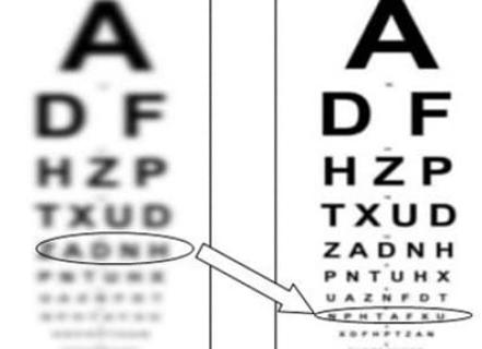
- Interocular difference in visual acuity (typically ≥2 lines on a crowded optotype chart).
- Reduced stereoacuity on random-dot or contour stereotests.
- Normal anterior/posterior segment exam.
- Cycloplegic refraction reveals anisometropia and/or high astigmatism.
- Cover tests may show tropia or phoria.
Strabismic amblyopia
- Constant, typically unilateral strabismus (often esotropia).
- Fixation preference for the nondeviating eye; suppression scotoma on binocular testing.
- Reduced stereopsis out of proportion to refractive findings.
Anisometropic amblyopia
- No manifest strabismus.
- Significant interocular spherical or cylindrical difference (eg, ≥1–1.5 D hyperopia or ≥1.5–2.0 D astigmatism commonly associated).
- Crowding phenomenon prominent; stereoacuity variably reduced.
Combined mechanism
- Features of both strabismus and anisometropia.
Deprivation amblyopia
- History/exam shows media opacity (eg, congenital cataract), severe ptosis obscuring the visual axis, corneal opacity, or vitreous hemorrhage.
- Most severe acuity loss; urgent management required to minimize permanent deficit.
Complications
- Persistent reduction in best-corrected acuity.
- Loss of stereopsis and binocular function affecting school, sports, and certain occupations.
- Increased lifetime risk if the better-seeing eye is injured or diseased; protective eyewear is recommended.
- Recurrence after successful therapy, particularly within the first year off treatment [6].
- Psychosocial impact (treatment adherence, self-image with patching).
Diagnosis
Clinical evaluation
- Age-appropriate monocular visual acuity testing (eg, Lea Symbols, HOTV, crowded optotypes; Teller acuity cards in infants).
- Ocular alignment assessment (cover–uncover, alternate cover, prism cover tests).
- Full anterior and posterior segment examination to exclude organic pathology (eg, cataract, maculopathy, optic neuropathy).
- Cycloplegic retinoscopy/refraction (eg, cyclopentolate 1%).
- Stereoacuity testing and suppression assessment (Worth 4-dot).
Diagnostic criterion: reduced best-corrected acuity in one or both eyes unexplained by structural abnormality, in the presence of a risk factor (strabismus, anisometropia, deprivation) and improved acuity with amblyopia therapy.
Imaging and laboratory testing
- Not routinely indicated.
- Optical coherence tomography (OCT) may be used to exclude subtle macular disease when clinical suspicion exists.
- Neuroimaging is reserved for atypical presentations (eg, severe unilateral loss without amblyogenic factors, afferent pupillary defect, neurologic signs).
- No laboratory tests are diagnostic for amblyopia.
Differential diagnosis
| Entity | Distinguishing features |
|---|---|
| Uncorrected bilateral refractive error | Symmetric decreased acuity; improves with appropriate refraction; normal binocular function when corrected |
| Congenital/developmental cataract | Lens opacity on slit-lamp; leukocoria; decreased red reflex; deprivation mechanism |
| Ptosis (severe) | Lid covers visual axis; improves with lid elevation; risk for deprivation amblyopia |
| Corneal opacity/scar (eg, after keratitis) | Corneal haze/scar on exam; history of ulcer or trauma; irregular astigmatism |
| Macular disease (eg, Best disease) | Abnormal macular reflex; OCT/ERG abnormalities; family history |
| Optic nerve hypoplasia/dysplasia | Small disc, double-ring sign; RAPD; associated endocrine/neurologic findings |
| Retinal dystrophies | Night blindness, color vision changes; abnormal ERG; bilateral involvement |
| Toxic/nutritional optic neuropathy | Symmetric central scotomas; dyschromatopsia; relevant exposures/diet |
| Nonorganic (functional) visual loss | Inconsistent findings; normal objective tests; variable performance |
Treatment
Goals: maximize best-corrected acuity in the amblyopic eye, restore binocular function when possible, and minimize recurrence.
Medical management
- Full refractive correction
- Prescribe full-time glasses or contact lenses to correct significant refractive error and anisometropia.
- Refractive adaptation alone can yield substantial improvement over weeks to months in anisometropic amblyopia [3].
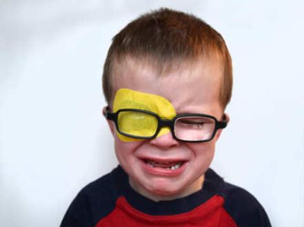
- Occlusion (patching) of the better-seeing eye
- Moderate amblyopia: 2 hours/day patching typically as effective as 6 hours/day when combined with near activities [3].
- Severe amblyopia: 6 hours/day often effective; full-time patching is rarely necessary and may increase adverse effects [4].
- Monitor every 6–12 weeks; adjust dose based on response and adherence.
- Adverse effects: skin irritation, social stigma, rare reverse amblyopia in the patched eye (mitigated by appropriate dosing and follow-up).
- Pharmacologic penalization with atropine 1% ophthalmic solution (off-label for amblyopia)
- Moderate amblyopia: 1 drop in the better-seeing eye daily or weekend-only dosing provides improvement comparable to patching in many children [4].
- Mechanism: cycloplegia and blur of the better-seeing eye at near and often distance in hyperopic eyes.
- Adverse effects: photophobia, blurred near vision, systemic anticholinergic effects (flushing, fever, dry mouth, tachycardia) are uncommon with ocular use; instruct caregivers on punctal occlusion and to seek care for systemic symptoms.
- Contraindications/cautions: known atropine hypersensitivity; use caution in children with underlying cardiac arrhythmias. Angle closure is rare in children.
- Bangerter filters (graded translucent foils applied to spectacle lens of the better-seeing eye)
- Useful alternative when patching/atropine adherence is poor; titratable density; generally well tolerated [7].
- Adjuncts and emerging therapies
- Binocular/video game–based treatments show mixed results and are not standard of care; consider only as adjuncts within clinical guidance [7].
- Pharmacologic agents such as levodopa or citicoline have inconsistent evidence and are not routinely recommended [7].
Adherence strategies:
- Educate caregivers on goals and timeline; integrate patching during screen-free near tasks (coloring, puzzles).
- Use reward systems; address bullying concerns with schools.
- Taper patching (eg, stepwise reduction) after success to reduce recurrence risk [6].
Procedural and surgical management
- Strabismus surgery
- Indicated to correct ocular alignment for binocular potential and psychosocial/cosmetic benefit.
- Does not replace amblyopia therapy; continue occlusion/penalization pre- and postoperatively per ophthalmologist.
- Deprivation causes
- Congenital cataract extraction in infancy (ideally within weeks for unilateral dense cataract) plus prompt optical correction (contact lens or intraocular lens as age-appropriate) and intensive amblyopia therapy [1,7].
- Surgical correction of severe ptosis that obscures the visual axis.
- Management of corneal opacities; treat underlying disease (eg, infectious keratitis) to clear the axis when possible.
Special populations
- Infants and toddlers
- Deprivation amblyopia is an emergency for visual development; expedited surgery/therapy is critical.
- Use preferential looking tests; frequent follow-up for rapid changes.
- Older children and adolescents
- Treatment can still improve acuity, especially if previously untreated; response is generally less robust and slower than in younger children [5].
- Neurodevelopmental disorders
- Expect greater challenges with testing and adherence; simplify regimens and engage multidisciplinary support.
- Adults
- Traditional therapies yield limited average gains; selected adults may benefit from targeted training, but evidence remains limited and such approaches are not standard [7].
- Pregnancy/lactation
- Typically not applicable because treatment targets the child; caregivers using atropine on the child should observe standard handling and hand hygiene.
Prognosis
Prognosis is best with early detection and treatment before age 7–8, anisometropic etiology, moderate baseline acuity loss, and good adherence. Many children gain several lines of acuity within 6–16 weeks of active therapy, followed by slower improvement. Deprivation amblyopia carries the poorest prognosis. Approximately one quarter of successfully treated children experience recurrence within the first year after stopping therapy; tapering and maintenance monitoring reduce risk [6].
Prevention and Patient Counseling
- Adhere to pediatric vision screening (ages 3–5) and earlier referral for suspected strabismus or visual axis obstruction [5].
- Ensure full-time wear of prescribed glasses.
- Supervise children during patching; encourage engaging near activities during occlusion.
- Protect the better-seeing eye with polycarbonate lenses during sports and high-risk activities.
- For eyelid lesions that cover the pupil in infants (eg, large chalazia) or any persistent ocular redness/pain, seek prompt evaluation to prevent deprivation; severe corneal infections can scar and impair vision.
For context on eye surface conditions sometimes confused with vision problems, see our resources on chalazion, bacterial conjunctivitis, and keratitis.
When to Seek Immediate Care
- Sudden vision loss in either eye.
- A droopy eyelid or eyelid mass covering the pupil in an infant or toddler.
- A red, painful eye with light sensitivity or reduced vision.
- New-onset constant eye turn with headache, vomiting, or other neurologic symptoms.
- Eye injury or chemical exposure.
- Worsening vision in the better-seeing eye.
References
- American Academy of Ophthalmology Pediatric Ophthalmology/Strabismus Panel. Amblyopia Preferred Practice Pattern. San Francisco, CA: AAO; 2022. https://www.aao.org/preferred-practice-pattern/amblyopia-ppp-2022.
- National Eye Institute. Facts About Amblyopia. National Institutes of Health. https://www.nei.nih.gov/learn-about-eye-health/eye-conditions-and-diseases/amblyopia. Accessed July 24, 2026.
- Pediatric Eye Disease Investigator Group (PEDIG). A randomized trial of patching regimens for treatment of moderate amblyopia in children. Arch Ophthalmol. 2003;121(5):603-611..
- Pediatric Eye Disease Investigator Group (PEDIG). A randomized trial of atropine vs patching for treatment of moderate amblyopia in children. N Engl J Med. 2002;347(9):621-629..
- US Preventive Services Task Force. Vision Screening in Children Aged 3 to 5 Years: US Preventive Services Task Force Recommendation Statement. JAMA. 2017;318(9):836-844. doi:10.1001/jama.2017.11260.
- Pediatric Eye Disease Investigator Group (PEDIG). Risk of amblyopia recurrence after cessation of treatment. J AAPOS. 2004;8(5):420-428..
- Wallace DK, Repka MX, Lee KA, et al. Amblyopia treatment outcomes and evidence-based options. Ophthalmology. 2018;125(9):1489-1503..
Disclaimer: This article is for informational purposes and is not a substitute for professional medical advice, diagnosis, or treatment. Always consult a qualified eye care professional about your specific condition. If you have sudden vision loss, severe eye pain, or an eye injury, seek emergency care immediately.