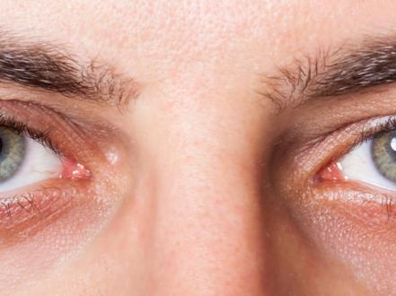
Bacterial conjunctivitis is acute or chronic inflammation of the bulbar and palpebral conjunctiva caused by pathogenic bacteria, characterized by mucopurulent discharge, conjunctival injection (redness), grittiness or foreign-body sensation, and eyelids matted on awakening. Diagnosis is clinical; cultures are reserved for severe, atypical, hyperacute, recurrent, or nonresponding cases. First-line treatment is topical antibiotics and supportive care; systemic therapy is required for gonococcal and chlamydial infections.

Key Points

  • Bacterial conjunctivitis causes purulent or mucopurulent discharge, eyelid crusting, and diffuse conjunctival redness; itching is less prominent than in allergic disease.
  • Contact lens wearers are at increased risk of corneal involvement, including Pseudomonas keratitis; they require urgent evaluation, lens cessation, and antipseudomonal coverage if infection is suspected.
  • Hyperacute conjunctivitis with copious purulent discharge suggests Neisseria gonorrhoeae and is an emergency requiring systemic ceftriaxone and STI evaluation per CDC guidelines.
  • Most uncomplicated cases are self-limited; topical antibiotics shorten symptom duration and hasten microbiologic cure modestly.
  • Pain, photophobia, or vision loss are atypical and suggest keratitis, uveitis, or angle-closure glaucoma rather than isolated conjunctivitis.
  • Avoid topical corticosteroids unless an ophthalmologist excludes herpetic keratitis and other corneal disease.

Anatomy and Physiology

The conjunctiva is a transparent mucous membrane lining the inner eyelids (palpebral conjunctiva) and reflecting onto the sclera (bulbar conjunctiva). It contains nonkeratinized epithelium with goblet cells, accessory lacrimal glands, and a rich vascular and lymphoid network. Normal tear film, blinking, and innate immunity (lysozyme, lactoferrin) protect against infection. Bacterial adherence and overgrowth trigger conjunctival hyperemia, papillary response, and exudation.

Etiology

Common pathogens vary by age and risk factors:

  • Children: nontypeable Haemophilus influenzae, Streptococcus pneumoniae, Moraxella catarrhalis [1].
  • Adults: Staphylococcus aureus (including MRSA), S. pneumoniae, H. influenzae [1,2].
  • Contact lens wearers: increased risk of Gram-negative organisms, notably Pseudomonas aeruginosa, often as keratitis rather than isolated conjunctivitis [2].
  • Hyperacute: Neisseria gonorrhoeae (gonococcal conjunctivitis) [3].
  • Chronic (>4 weeks) or adult inclusion conjunctivitis: Chlamydia trachomatis (serovars D–K), sexually transmitted [3,4].

Predisposing factors include poor hand hygiene, close contact exposures, concurrent blepharitis, chronic dacryocystitis, ocular surface disease (eg, dry eye), and contaminated cosmetics or lenses.

Pathophysiology

Bacteria colonize the conjunctival epithelium and lid margins, overcoming tear film defenses and mechanical clearance. The host inflammatory response increases vascular permeability and neutrophil influx, producing mucopurulent discharge and papillary hypertrophy. Hyperacute gonococcal infection penetrates intact epithelium and can rapidly involve the cornea, risking ulceration and perforation. Chlamydia causes a chronic follicular response with superior pannus in some cases.

Epidemiology

Bacterial conjunctivitis is among the most common ocular infections in primary and urgent care. In children, bacterial etiologies predominate; in adults, viral causes are more frequent while bacterial conjunctivitis remains common [1,2]. Exact US incidence varies by setting and season, with increased rates in daycare/schools and during winter months in temperate climates.

Classification

  • Acute bacterial conjunctivitis (typical mucopurulent form)
  • Hyperacute bacterial conjunctivitis (gonococcal)
  • Chronic bacterial conjunctivitis (>4 weeks), often chlamydial; can also reflect staphylococcal blepharoconjunctivitis
  • Special contexts:
    • Contact lens–associated red eye (evaluate for keratitis)
    • Postoperative or immunocompromised host infections

Symptoms and Signs

Acute bacterial conjunctivitis (typical)

  • Rapid onset redness in one eye, often second eye involvement within 24–48 hours
  • Mucopurulent discharge, eyelids stuck on awakening
  • Grittiness or foreign-body sensation, tearing
  • Diffuse bulbar and palpebral injection with papillary reaction
  • Mild eyelid edema; minimal pain; no photophobia; vision normal after clearing discharge
  • Preauricular lymphadenopathy usually absent

Hyperacute (gonococcal) conjunctivitis

  • Abrupt onset with copious, thick purulent discharge reaccumulating after wiping
  • Marked chemosis (conjunctival swelling), severe eyelid edema, tenderness
  • Preauricular lymphadenopathy may be present
  • Corneal involvement common; risk of ulceration and perforation
  • Often concomitant urogenital symptoms or risk factors for STIs

Chronic bacterial (chlamydial) conjunctivitis

  • Weeks to months of unilateral or bilateral redness and discharge
  • Mucopurulent discharge, prominent follicles on inferior palpebral conjunctiva
  • Minimal itching; variable photophobia
  • Superior corneal pannus and micropannus may be seen
  • Associated urethritis/cervicitis; sexual partners often infected

Complications

  • Keratitis and corneal ulceration (notably Pseudomonas in lens wearers; gonococcal)
  • Preseptal cellulitis
  • Dacryocystitis
  • Reactive subepithelial infiltrates
  • Chronic blepharoconjunctivitis
  • Rare: corneal perforation (hyperacute gonococcal), vision loss

Diagnosis

Clinical evaluation

  • History: onset, discharge character, laterality, contact lens use, trauma/chemical exposure, prior similar episodes, systemic STI risks, associated pain/photophobia/blurred vision.
  • Examination: visual acuity, external inspection, conjunctival injection pattern, papillae vs follicles, discharge amount/quality, preauricular nodes, corneal clarity with fluorescein staining and cobalt blue light.
  • Remove and inspect contact lenses; do not reinsert until resolution.

Indications to obtain microbiologic testing:

  • Hyperacute presentation (suspect N. gonorrhoeae)
  • Chronic conjunctivitis (suspect C. trachomatis) or recurrent disease
  • Severe disease, corneal involvement, immunocompromised host, neonatal disease
  • Postoperative infection or poor response to appropriate empiric therapy

Imaging and laboratory testing

  • Conjunctival Gram stain and culture on appropriate media (eg, Thayer-Martin for Neisseria) for hyperacute cases [3].
  • Nucleic acid amplification test (NAAT) for C. trachomatis from conjunctival swab when chronic follicular conjunctivitis suspected; test for concurrent genital infection [3,4].
  • No imaging is indicated for uncomplicated cases.

Differential diagnosis

Entity Distinguishing features
Viral conjunctivitis (adenoviral) Watery discharge, prominent follicular reaction, preauricular nodes, often starts unilateral then bilateral; recent URI; subepithelial corneal infiltrates possible
Allergic conjunctivitis Intense itching, stringy/ropy discharge, atopy history, chemosis; seasonal/perennial pattern; minimal crusting
Keratitis (bacterial/viral) Pain, photophobia, decreased vision; focal corneal opacity/stain; urgent referral see our keratitis guide: https://myvisioncare.org/disease/keratitis
Anterior uveitis Photophobia, ciliary flush, consensual pain, cells/flare in anterior chamber
Blepharitis Lid margin inflammation, collarettes, meibomian dysfunction; symptoms worse on awakening; may coexist with conjunctivitis
Dry eye disease Burning, fluctuating vision, worsens later in day; minimal discharge see dry eye overview: https://myvisioncare.org/blog/dry-eye-syndrome
Episcleritis Sectoral redness, mild tenderness, normal discharge, self-limited
Dacryocystitis Painful medial canthus swelling, reflux of pus from punctum; systemic antibiotics indicated
Chalazion/hordeolum Focal lid nodule or tender pustule; localized rather than diffuse conjunctival involvement learn about chalazion: https://myvisioncare.org/disease/chalazion
Chemical/toxic conjunctivitis Exposure history (irritants, preservatives), burning, stringy discharge, punctate keratitis

Treatment

Medical management

Supportive care for all patients:

  • Discontinue contact lenses; discard or disinfect lenses and cases before reuse.
  • Warm compresses to remove crusts; cold compresses for comfort; preservative-free artificial tears.
  • Hand hygiene; avoid sharing towels/cosmetics; clean eyelid margins.

Topical antibiotics (typical acute bacterial conjunctivitis):

  • Erythromycin ophthalmic ointment 0.5-inch ribbon to affected eye(s) qid (4 times daily) for 7 days [1,2].
  • Trimethoprim–polymyxin B drops: 1 drop q3h while awake (up to 6 times daily) for 7 days; many clinicians use qid (4 times daily) for 5–7 days [1,2].
  • Alternative agents (reserve for contact lens wearers, severe disease, or intolerance; cover Gram-negatives including Pseudomonas when keratitis risk is present):
    • Ofloxacin 0.3%: 1–2 drops q2–4h for 2 days, then qid to complete 7 days.
    • Ciprofloxacin 0.3%: 1–2 drops q2h while awake for 2 days, then q4h to complete 7 days.
    • Moxifloxacin 0.5%: 1 drop tid (3 times daily) for 7 days.
    • Gatifloxacin 0.5%: 1 drop qid for 7 days.
    • Besifloxacin 0.6%: 1 drop tid for 7 days.
      Notes: Fluoroquinolones are preferred if corneal involvement is suspected, particularly in contact lens wearers [1,2]. Avoid neomycin-containing products due to hypersensitivity risk.

Gonococcal conjunctivitis (hyperacute) — emergency management (adults and adolescents):

  • Ceftriaxone 1 g IM in a single dose plus saline eye irrigation; evaluate/treat for concurrent STIs per CDC; add antichlamydial therapy if chlamydial coinfection not excluded [3]. Topical antibiotics are adjunctive only.
  • Hospitalize if corneal involvement or poor follow-up; urgent ophthalmology referral.

Chlamydial adult inclusion conjunctivitis:

  • Azithromycin 1 g PO single dose or doxycycline 100 mg PO bid for 7 days; treat sexual partners; coordinate urogenital testing [3,4]. Topical antibiotics are optional adjuncts.

Analgesia:

  • Oral acetaminophen or NSAIDs as needed. Avoid topical anesthetics outside clinical settings.

Corticosteroids:

  • Avoid topical steroids in suspected infectious conjunctivitis unless directed by an ophthalmologist after corneal disease and herpetic infection are excluded.

Antibiotic stewardship:

  • Uncomplicated, mild cases may resolve without antibiotics; shared decision-making is appropriate in non–contact lens adults with reliable follow-up [5]. Start antibiotics promptly if symptoms worsen or fail to improve in 1–2 days.

Procedural and surgical management

  • No routine procedures are indicated. Copious saline irrigation can aid discharge removal in hyperacute cases.
  • Manage associated blepharitis with lid hygiene; consider in-office expression for meibomian gland dysfunction if chronic.

Special populations

  • Pediatric: Erythromycin ointment or trimethoprim–polymyxin B drops are first-line. Topical fluoroquinolones may be used when corneal involvement is suspected or for contact lens wearers. Neonatal conjunctivitis requires separate evaluation and systemic therapy pathways.
  • Pregnancy/lactation: Erythromycin ointment and trimethoprim–polymyxin B drops are generally acceptable due to minimal systemic absorption; avoid systemic doxycycline. For chlamydia in pregnancy, azithromycin 1 g PO single dose is recommended [3]. During lactation, topical agents have low risk.
  • Immunocompromised/ocular surface disease: Lower threshold for cultures; monitor closely for keratitis and atypical organisms.

Prognosis

With appropriate care, typical bacterial conjunctivitis improves within 24–48 hours of starting topical antibiotics and resolves in 5–7 days. Without antibiotics, many cases resolve over 7–10 days [5]. Hyperacute gonococcal disease can threaten the cornea within hours to days. Vision loss is rare and usually reflects corneal involvement.

Prevention and Patient Counseling

  • Hand hygiene, avoid eye rubbing, and do not share towels, cosmetics, or eye drops.
  • Stop contact lenses during infection; replace or disinfect lenses and cases before reuse; avoid swimming and hot tubs with lenses.
  • Clean eyelid margins; manage chronic blepharitis and dry eye to reduce recurrence.
  • Household spread risk exists; avoid close contact until discharge subsides; some institutions require 24 hours of antibiotics before return to group settings; policies vary.

When to Seek Immediate Care

  • Severe eye pain, light sensitivity, or sudden vision changes
  • Contact lens wearer with a red, painful, or photophobic eye
  • Copious thick pus with rapid worsening (possible gonococcal infection)
  • Eye trauma or chemical splash
  • Marked eyelid swelling, fever, or facial redness
  • Inability to open the eye or concern for corneal ulcer
  • Eye discharge in an infant under 1 month old

References

  1. American Academy of Ophthalmology Cornea/External Disease Panel. Conjunctivitis Preferred Practice Pattern. AAO; 2023. https://www.aao.org/ppp
  2. Azari AA, Barney NP. Conjunctivitis: A Systematic Review of Diagnosis and Treatment. JAMA. 2013;310(16):1721-1729. doi:10.1001/jama.2013.280318. [Review; still widely cited]
  3. CDC. Sexually Transmitted Infections Treatment Guidelines, 2021: Gonococcal Infections and Chlamydial Infections. https://www.cdc.gov/std/treatment-guidelines/default.htm
  4. Darougar S, Forsey T, Jones BR. Chlamydial conjunctivitis. Int Ophthalmol Clin. 1986;26(3):73-86.
  5. Shekhawat NS, Shtein RM, Blachley TS, Stein JD. Antibiotic Prescription Fills for Acute Conjunctivitis among Enrollees in a Large United States Managed Care Network. Ophthalmology. 2017;124(8):1099-1107. doi:10.1016/j.ophtha.2017.02.021.
  6. Cochrane Eyes and Vision Group. Antibiotics versus placebo for acute bacterial conjunctivitis. Cochrane Database Syst Rev. 2022;.
  7. NEI (National Eye Institute). Conjunctivitis (Pink Eye). https://www.nei.nih.gov/learn-about-eye-health/eye-conditions-and-diseases/conjunctivitis
  8. StatPearls. Bacterial Conjunctivitis. Updated 2024. https://www.ncbi.nlm.nih.gov/books/NBK546683/

This article is for informational purposes and is not a substitute for professional medical advice, diagnosis, or treatment. Always consult a qualified eye care professional about your specific condition. If you have sudden vision loss, severe eye pain, or an eye injury, seek emergency care immediately.