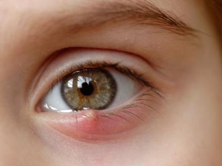
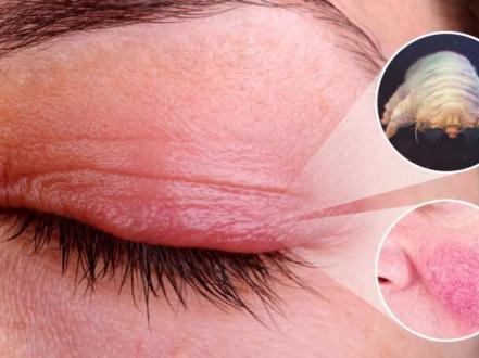
A chalazion is a sterile, chronic, lipogranulomatous inflammation caused by obstruction of a meibomian (tarsal) or, less often, Zeis gland in the eyelid. It presents as a firm, typically painless eyelid nodule that may induce localized eyelid swelling and, if large, corneal astigmatism with blurred vision. Diagnosis is clinical by external and everted eyelid examination. Initial management is warm compresses and lid hygiene; persistent lesions are treated with intralesional corticosteroid injection or incision and curettage.
Key Points
- Chalazion is a noninfectious granulomatous reaction to retained meibum from an obstructed eyelid oil gland.
- Cardinal findings: firm, non-tender eyelid nodule; overlying erythema may be present early; pain is minimal unless there is secondary inflammation.
- Risk factors include blepharitis, meibomian gland dysfunction, acne rosacea, and prior chalazion.
- Differentiate from hordeolum (stye), preseptal cellulitis, and sebaceous gland carcinoma; biopsy atypical, recurrent, or nonresolving lesions.
- Most resolve with conservative care; persistent lesions respond to intralesional triamcinolone or incision and curettage.
- Prompt care is needed for rapidly worsening pain/redness, fever, vision changes, or recurrent lesions at the same site in older adults.
Anatomy and Physiology
Meibomian glands are modified sebaceous glands embedded vertically within the tarsal plates of the eyelids; they secrete meibum, the lipid layer of the tear film that reduces evaporation. Glands of Zeis are small sebaceous glands associated with eyelashes at the lid margin. Ductal obstruction leads to retention of lipid material, acinar rupture, and a granulomatous foreign body response in the tarsus or lid margin.
Etiology
- Meibomian gland duct obstruction with retained lipid
- Preexisting lid margin disease:
- Chronic anterior/posterior blepharitis
- Meibomian gland dysfunction (MGD)
- Acne rosacea and seborrheic dermatitis
- Contributing factors: poor lid hygiene, Demodex infestation, prior chalazion, frequent eye rubbing, oil-based cosmetics
Pathophysiology
Ductal blockage causes stasis of meibum and intraglandular pressure. Glandular rupture extravasates lipid into surrounding stroma, inciting a lipogranulomatous reaction with epithelioid histiocytes, multinucleated giant cells, and chronic inflammatory cells. The resultant mass is well circumscribed, often within the tarsus (internal variant). Secondary bacterial colonization is uncommon; chalazion is not primarily infectious.
Epidemiology
Chalazion is among the most common eyelid lesions encountered in primary eye care and oculoplastics clinics. All ages are affected, with peaks in adolescents/young adults and patients with rosacea/MGD. Bilateral or multiple lesions occur in lid margin disease. Exact US incidence and prevalence are not well established.
Classification
- By gland of origin:
- Internal chalazion: meibomian gland (tarsal)
- External chalazion: Zeis gland (lid margin; less common)
- By course:
- Acute inflammatory phase versus chronic organized nodule
- Primary versus recurrent (same location)
- By clinical concern:
- Typical versus atypical (recurrent, madarosis, ulceration, or older adult—raise concern for malignancy)
Symptoms and Signs
-
Typical chalazion
- Symptoms: painless eyelid lump, mild localized tenderness early, cosmetic concern; foreign body sensation or tearing may occur.
- Signs: firm, localized, nonfluctuant nodule within tarsus or at lid margin; minimal erythema; conjunctival surface may show a yellow-gray granuloma on lid eversion; associated telangiectatic lid margin and meibum inspissation in MGD.
- Large lesions can indent the cornea, causing induced astigmatism and transient blurred vision.
-
Acute inflammatory phase
- Symptoms: focal lid tenderness, warmth, localized erythema; pain less than a hordeolum.
- Signs: erythematous swelling centered over tarsus; no diffuse cellulitis.
-
Recurrent or atypical lesions
- Symptoms: repeated nodules at same site, persistent firm plaque, lash loss.
- Signs: focal thickening or ulceration at margin, yellowish infiltration, madarosis; consider sebaceous gland carcinoma.
Complications
- Induced corneal astigmatism and visual blur; in infants/young children, risk of deprivation or astigmatic amblyopia if large and persistent
- Pyogenic granuloma formation on palpebral conjunctiva
- Secondary infection (preseptal cellulitis) is uncommon
- Skin depigmentation or fat atrophy after intralesional steroid (procedure-related)
- Recurrence, especially with untreated blepharitis/MGD or rosacea
Diagnosis
Clinical evaluation
- History: onset, pain severity, prior similar lesions, rosacea/blepharitis, cosmetic use, contact lens wear, recurrence at same site.
- Examination:
- External inspection and palpation for a firm, non-tender tarsal nodule.
- Lid eversion: inspect palpebral conjunctiva for granuloma; assess for pyogenic granuloma.
- Lid margin: look for MGD (plugged orifices, turbid meibum), collarettes, telangiectasias.
- Cornea: assess curvature/regularity and surface staining if visual symptoms.
- Red flags: lash loss, ulceration, persistent induration, regional lymphadenopathy.
Imaging and laboratory testing
Routine imaging and laboratory tests are not indicated. Send tissue for histopathology in recurrent, atypical, or nonresolving lesions, especially in older adults, to exclude sebaceous gland carcinoma or other malignancy. Imaging (eg, orbital CT/MRI) is reserved for suspected orbital disease, which is not typical of chalazion.
Differential diagnosis
| Entity | Distinguishing features |
|---|---|
| Hordeolum (stye) | Acute, painful, tender, erythematous pustule at lash follicle (external) or meibomian gland (internal); often points and drains; infectious (usually Staphylococcus). |
| Preseptal cellulitis | Diffuse, tender eyelid edema/erythema, warmth, sometimes fever; no focal tarsal nodule; normal ocular motility and globe. |
| Sebaceous gland carcinoma | Recurrent or nonresolving firm lesion; yellowish thickening, madarosis, margin ulceration; pagetoid spread; older adults; requires biopsy. |
| Basal cell carcinoma | Pearly, telangiectatic, ulcerated or indurated lid lesion; slow growth; lower lid common; loss of lashes over lesion. |
| Epidermal inclusion cyst | Mobile, subcutaneous, dome-shaped lesion; central punctum; not tarsal; usually noninflammatory unless ruptured. |
| Pyogenic granuloma | Rapidly growing, friable red mass on conjunctiva or skin after surgery/trauma/chalazion; bleeds easily. |
| Dacryocystitis | Painful, erythematous swelling inferomedial to medial canthus over lacrimal sac; epiphora; purulent reflux from punctum with pressure. |
| Conjunctival/lid lymphoma | Salmon-colored conjunctival patch; firm, painless lid mass; often insidious; requires biopsy. |
Treatment
Medical management
- Conservative care (first-line for most)
- Warm compresses: apply for 10–15 minutes, 3–6 times daily, followed by gentle lid massage toward the lid margin to express meibum [1,2].
- Lid hygiene: daily lid scrubs (commercial lid wipes or diluted nonirritating cleanser) and management of underlying blepharitis/MGD.
- Topical antibiotics: not effective for the sterile nodule; consider erythromycin ophthalmic ointment 0.5% bid–qid to lid margins for concurrent anterior blepharitis (5–7 days) [1,3].
- Systemic antibiotics (off-label for chalazion; useful for associated MGD/rosacea or recurrent lesions)
- Doxycycline 50–100 mg PO qday–bid for 2–4 weeks, then taper to 20–50 mg qday for maintenance in recalcitrant MGD/rosacea [1,3]. Avoid in pregnancy, breastfeeding, and children <8 years.
- Alternatives when tetracyclines are contraindicated: azithromycin PO pulse regimens or erythromycin PO may be considered for rosacea/MGD.
- Intralesional corticosteroid injection (office-based)
- Triamcinolone acetonide (TA) 5–10 mg/mL, 0.1–0.3 mL injected transconjunctivally into the lesion; may repeat after 2–6 weeks if residual [1,2,4].
- Adverse effects: skin depigmentation (higher risk with transcutaneous injection and darker skin), subcutaneous fat atrophy, steroid-related intraocular pressure rise if inadvertently intraocular, globe perforation, retinal vascular occlusion from inadvertent intravascular injection (rare) [4].
- Comparable resolution rates to incision and curettage in randomized trials.
Procedural and surgical management
- Incision and curettage (I&C)
- Indications: failure of conservative care, large or visually significant lesion, patient preference, or when tissue diagnosis is needed.
- Technique: local anesthesia; chalazion clamp; transconjunctival vertical tarsal incision; curettage of granulomatous contents; hemostasis; optional intralesional TA at completion; antibiotic ointment to incision [1,2].
- Send tissue for histopathology if lesion is recurrent, atypical, or in older adults.
- CPT coding: incision and curettage of chalazion—single or multiple lesions.
- Pediatric considerations: I&C often requires sedation or general anesthesia in young children; balance anesthesia risks with potential for amblyopia from large lesions.
Special populations
- Pediatric
- Large lesions can induce significant astigmatism and risk amblyopia; consider earlier procedural management if visual axis is affected.
- Avoid tetracyclines in children <8 years; use erythromycin ophthalmic ointment for concurrent blepharitis as needed.
- Pregnancy and lactation
- Prefer conservative measures. Avoid tetracyclines; erythromycin ophthalmic ointment is acceptable. Defer intralesional steroid if possible; if required, use the lowest effective dose with obstetric input.
- Immunocompromised
- Maintain a low threshold for culture/biopsy of atypical, draining, or nonhealing lesions to exclude atypical infection or neoplasm.
Prognosis
Most chalazia resolve over weeks to months with conservative therapy. Intralesional TA or I&C provides high rates of lesion resolution when needed. Recurrence is common in patients with untreated MGD or rosacea. Visual prognosis is excellent; induced astigmatism is reversible after lesion resolution. Delay in treating large lesions in infants can lead to amblyopia.
Prevention and Patient Counseling
- Daily lid hygiene: warm compresses and gentle lid scrubs to maintain meibomian gland function.
- Manage underlying blepharitis/MGD and rosacea to reduce recurrence.
- Avoid eye rubbing; remove eye makeup thoroughly; replace old eye cosmetics.
- Chalazion is not contagious.
- Persistent, recurrent, or atypical lesions warrant evaluation and possible biopsy to exclude malignancy.
When to Seek Immediate Care
- Sudden vision loss, double vision, severe eye pain, or inability to move the eye
- Rapidly worsening eyelid redness, swelling, warmth, or fever
- Diffuse eyelid infection signs (swelling spreading beyond a focal lump)
- A chalazion that recurs in the same spot, especially in an older adult
- New lash loss or a nonhealing ulcer at the eyelid margin
- Severe headache, nausea, or vomiting after an eye injection
References
- American Academy of Ophthalmology EyeWiki. Chalazion. https://eyewiki.aao.org/Chalazion. Accessed July 24, 2026.
- StatPearls. Chalazion. StatPearls Publishing; updated 2024. https://www.ncbi.nlm.nih.gov/books/NBK499889/. Accessed July 24, 2026.
- American Academy of Ophthalmology Preferred Practice Pattern. Blepharitis PPP. https://www.aao.org/preferred-practice-pattern/blepharitis-ppp. Accessed July 24, 2026.
- DailyMed. Triamcinolone acetonide injectable suspension, USP. https://dailymed.nlm.nih.gov/dailymed/. Accessed July 24, 2026.
- AAPOS. Chalazion. American Association for Pediatric Ophthalmology and Strabismus. https://aapos.org/glossary/chalazion. Accessed July 24, 2026.
- Cleveland Clinic. Chalazion: Symptoms, Causes & Treatment. https://my.clevelandclinic.org/health/diseases/17657-chalazion. Accessed July 24, 2026.
- Goawalla A, Lee V. A prospective randomized treatment study comparing three treatment options for chalazia. Eye (Lond). 2007;21(6): (pages).
- Ben Simon GJ, et al. Intralesional triamcinolone vs incision and curettage for primary chalazia. Ophthalmology. 2011;118(12): (pages).
Disclaimer: This article is for informational purposes and is not a substitute for professional medical advice, diagnosis, or treatment. Always consult a qualified eye care professional about your specific condition. If you have sudden vision loss, severe eye pain, or an eye injury, seek emergency care immediately.