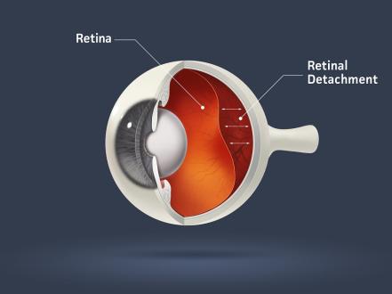
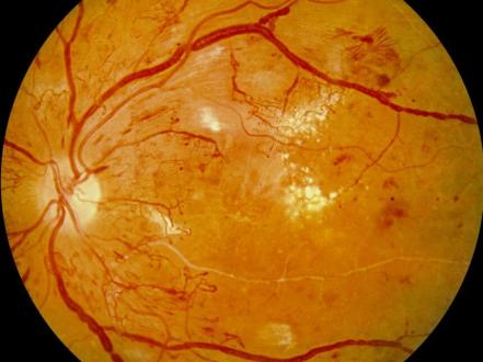
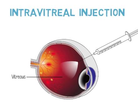
Diabetic retinopathy is a microvascular complication of diabetes mellitus that damages retinal capillaries and can cause vision loss through macular edema, ischemia, and pathologic neovascularization. Symptoms are often absent until advanced disease; patients may develop blurred vision, floaters, or sudden vision loss from vitreous hemorrhage or tractional retinal detachment. Diagnosis is by dilated fundus examination supported by optical coherence tomography (OCT) and fluorescein angiography. Treatment includes systemic risk-factor control, intravitreal anti–vascular endothelial growth factor (anti-VEGF) therapy for diabetic macular edema (DME) and some proliferative cases, focal/grid laser for select edema, panretinal photocoagulation (PRP) for proliferative diabetic retinopathy (PDR), and vitreoretinal surgery for complications.
Key Points
- Diabetic retinopathy is the leading cause of new blindness in working-age adults in the US; risk increases with diabetes duration and poor glycemic/blood pressure control [1,2].
- Most patients are asymptomatic until moderate-to-severe nonproliferative or proliferative disease; annual dilated retinal screening is essential (ADA/AAO) [1,2].
- Anti-VEGF therapy is first-line for center-involved DME; PRP remains standard for high-risk PDR, with anti-VEGF a validated alternative in selected patients [3–6].
- Intensive systemic management (A1c, blood pressure, lipids) slows onset and progression; fenofibrate reduces retinopathy progression in some patients but is not FDA-approved for this indication [1,2].
- Pregnancy may accelerate retinopathy; women with preexisting diabetes require eye exams before conception or in the first trimester and periodic follow-up [1].
- Aspirin for cardiovascular prevention is not contraindicated by retinopathy and does not increase ocular bleeding risk per existing evidence [1].
Anatomy and Physiology
The retina is a neurosensory tissue supplied by a dual circulation: inner retina by the retinal arterioles/capillaries and outer retina by the choroid. The inner blood–retina barrier (endothelial tight junctions and pericytes) maintains fluid homeostasis. VEGF is a hypoxia-inducible cytokine that increases vascular permeability and drives neovascularization. The macula subserves central vision; edema or ischemia here degrades visual acuity and contrast sensitivity.
Etiology
- Chronic hyperglycemia (type 1 or type 2 diabetes)
- Hypertension
- Dyslipidemia (elevated triglycerides, LDL)
- Duration of diabetes (major determinant)
- Nephropathy/chronic kidney disease
- Pregnancy (preexisting diabetes)
- Others: obstructive sleep apnea, smoking, anemia
Pathophysiology
Hyperglycemia triggers capillary basement membrane thickening, loss of pericytes, endothelial dysfunction, and microaneurysm formation. Capillary nonperfusion and ischemia ensue, leading to upregulation of VEGF and other cytokines. Consequences include:
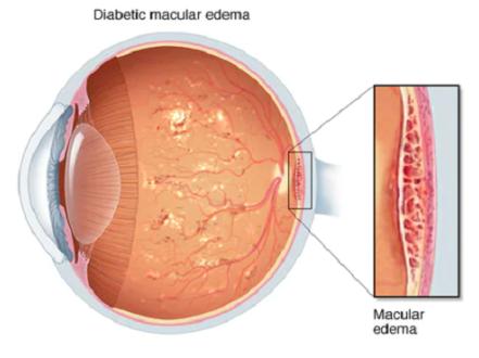
- Increased vascular permeability and leakage causing DME.
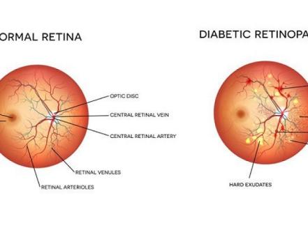
- Intraretinal hemorrhages, hard exudates (lipid), venous beading, and intraretinal microvascular abnormalities (IRMAs) in nonproliferative diabetic retinopathy (NPDR).
- Retinal neovascularization at the disc or elsewhere (NVD/NVE) in proliferative disease, with fragile vessels prone to vitreous/preretinal hemorrhage, fibrovascular proliferation, tractional retinal detachment, and neovascular glaucoma.
Epidemiology
- Approximately one-third of adults with diabetes have signs of diabetic retinopathy, and a smaller subset have vision-threatening disease (PDR or DME) [2,7].
- Diabetic retinopathy prevalence and severity correlate with diabetes duration; risk accumulates after 5–10 years and is highest beyond 20 years [1,2].
- In the US, diabetic retinopathy is a major cause of vision loss in working-age adults [1,7].
Classification
- Nonproliferative diabetic retinopathy (NPDR)
- Mild: microaneurysms only.
- Moderate: more than microaneurysms but not severe.
- Severe: “4-2-1 rule” (ETDRS): ≥20 intraretinal hemorrhages in 4 quadrants; venous beading in ≥2 quadrants; or IRMA in ≥1 quadrant (any one).
- Proliferative diabetic retinopathy (PDR): neovascularization of disc (NVD) or elsewhere (NVE), and/or vitreous/preretinal hemorrhage; “high-risk” PDR defined by ETDRS criteria (e.g., NVD ≥1/4–1/3 disc area or any NVD/NVE with vitreous hemorrhage).
- Diabetic macular edema (DME)
- Center-involved (CI-DME): edema involving the foveal center on OCT; primary determinant for intravitreal therapy.
- Non–center-involved DME: edema sparing the foveal center; often observed or treated with focal/grid laser.
Symptoms and Signs
- NPDR (mild to moderate)
- Symptoms: often none; intermittent mild blur.
- Signs: microaneurysms, dot-blot hemorrhages, hard exudates, cotton-wool spots (nerve fiber layer infarcts), mild venous changes.
- Severe NPDR
- Symptoms: reduced contrast sensitivity, fluctuating blur.
- Signs: numerous hemorrhages in all quadrants, venous beading, IRMAs, capillary nonperfusion on angiography.
- PDR
- Symptoms: new floaters, photopsias, sudden painless vision loss with vitreous hemorrhage; progressive field loss if untreated.
- Signs: NVD/NVE, preretinal/vitreous hemorrhage, fibrovascular proliferation, tractional retinal detachment; neovascularization of iris/angle in advanced disease.
- DME
- Symptoms: central blur, metamorphopsia (distorted lines), decreased reading speed; often chronic and fluctuating.
- Signs: retinal thickening on OCT, cystic changes, subretinal fluid; lipid exudates near the macula.
Complications
- Vision-threatening DME and/or macular ischemia
- Vitreous/preretinal hemorrhage
- Tractional retinal detachment (macula-on or macula-off)
- Combined tractional–rhegmatogenous retinal detachment
- Neovascular glaucoma
- Persistent visual dysfunction (contrast sensitivity, color vision, night vision impairment) even after edema resolves
Diagnosis
Clinical evaluation
- History: diabetes type and duration, A1c trajectory, blood pressure, lipid disorders, renal disease, pregnancy status, prior ocular treatments (laser, injections, surgery), visual symptoms (onset, laterality, floaters, scotomas).
- Examination
- Best-corrected visual acuity (ETDRS or Snellen), intraocular pressure.
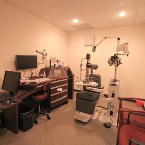
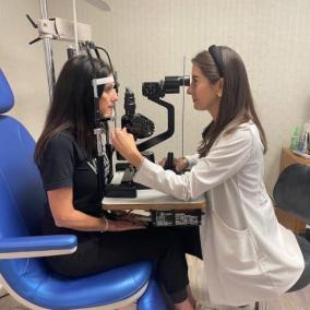
- Slit-lamp biomicroscopy with dilated fundus examination.
- Staging per ETDRS/AAO criteria.
- Document with color fundus photography; ultra-widefield imaging enhances peripheral assessment.
Imaging and laboratory testing
- OCT: quantifies retinal thickness, identifies cystoid spaces, subretinal fluid, and vitreoretinal interface pathology; essential for diagnosing and monitoring DME.
- Fluorescein angiography: maps areas of capillary nonperfusion, microaneurysms, leakage, and neovascularization; guides focal/grid laser and PRP.
- OCT angiography (OCT-A): noninvasive assessment of macular perfusion and neovascular complexes; complements, but does not replace, fluorescein angiography.
- B-scan ultrasonography: indicated for media opacity (dense vitreous hemorrhage) to exclude retinal detachment.
- Systemic labs (ordered/monitored by primary care/endocrinology): A1c, fasting lipid profile, renal function (eGFR, albuminuria) to guide risk-factor control.
Differential diagnosis
| Entity | Distinguishing features |
|---|---|
| Retinal vein occlusion | Unilateral; sudden vision loss; diffuse hemorrhages and cotton-wool spots along a venous distribution; dilated tortuous veins; risk factors include hypertension, glaucoma. |
| Hypertensive retinopathy | Arteriolar narrowing, AV nicking, flame hemorrhages; macular star; correlates with systemic hypertension rather than diabetes duration. |
| Ocular ischemic syndrome | Mid-peripheral hemorrhages, dilated but not tortuous veins, neovascularization; carotid stenosis; delayed choroidal/arterial filling on angiography; ocular/neck bruits. |
| Sickle cell retinopathy | Sea-fan neovascularization in periphery; history of hemoglobinopathy; salmon patches; younger age. |
| Radiation retinopathy | History of ocular/periocular radiation; telangiectasia and microaneurysms mimicking NPDR; latency months to years. |
| Inflammatory retinal vasculitis | Perivascular sheathing, vitreous cells; systemic autoimmune/infectious clues; responds to immunosuppression. |
Treatment
Goals are to prevent vision-threatening complications, reduce edema, regress neovascularization, and optimize visual function while coordinating systemic disease control.
Medical management
- Systemic optimization (in collaboration with primary care/endocrinology)
- Glycemic control: target A1c individualized; tighter control reduces onset and progression of retinopathy (DCCT/EDIC; UKPDS) [1,2].
- Blood pressure control: reduces retinopathy progression; ADA/ACC/AHA targets individualized (often <130/80 mm Hg) [1].
- Lipid management: statins per cardiovascular risk; fenofibrate has shown reduced progression in some studies (FIELD, ACCORD Eye) but lacks an FDA indication for retinopathy [1,2].
- Smoking cessation, weight management, sleep apnea evaluation when indicated.
- Aspirin for cardiovascular prevention may be continued; it does not worsen ocular bleeding in diabetic retinopathy [1].
- DME (center-involved) first-line intravitreal anti-VEGF therapy [3–6]
- Aflibercept 2 mg intravitreal every 4 weeks for 5 doses, then every 8 weeks; treat-and-extend common in practice (Eylea) [4].
- Ranibizumab 0.3 mg intravitreal monthly initially; interval extension per response (Lucentis 0.3 mg formulation for DME) [3].
- Faricimab 6 mg intravitreal every 4 weeks for 4 doses, then extend to every 8–16 weeks based on disease activity (Vabysmo) [5].
- Bevacizumab 1.25 mg intravitreal every 4–8 weeks is widely used off-label; randomized trials show variable relative efficacy versus labeled agents depending on baseline vision (DRCR Protocol T) [6]. Off-label use.
- Corticosteroids (consider in pseudophakic eyes or anti-VEGF nonresponders; monitor intraocular pressure)
- Dexamethasone intravitreal implant 0.7 mg (Ozurdex) typically every 3–4 months as needed; FDA-approved for DME [8].
- Fluocinolone acetonide 0.19 mg implant (Iluvien) releases ~0.2 µg/day up to 36 months; FDA-approved for DME in patients previously treated with corticosteroids and no significant IOP rise [9].
- Intravitreal triamcinolone acetonide (1–4 mg) is off-label for DME.
- Observation
- Non–center-involved DME and mild NPDR may be observed with systemic optimization and close follow-up.
Procedural and surgical management
- Focal/grid laser photocoagulation
- Indications: non–center-involved DME threatening the center; focal leakage from microaneurysms; adjunct to anti-VEGF in refractory cases (ETDRS-guided).
- Panretinal photocoagulation (PRP)
- Indications: high-risk PDR; may be used for severe NPDR with high risk of progression when follow-up is unreliable.
- Outcomes: reduces severe vision loss; may reduce peripheral field and dark adaptation; macular edema may transiently worsen [1].
- Anti-VEGF for PDR
- Ranibizumab demonstrated noninferiority to PRP for visual outcomes with less field loss but more visits/injections (Protocol S); appropriate in patients who can adhere to frequent follow-up, and when avoiding PRP side effects is desirable [10].
- Pars plana vitrectomy
- Indications: nonclearing vitreous hemorrhage, tractional retinal detachment threatening/affecting the macula, combined tractional–rhegmatogenous detachment, dense premacular hemorrhage.
- Preoperative intravitreal anti-VEGF (3–7 days before surgery) can reduce intraoperative bleeding but requires timely surgery to mitigate progression of traction.
Special populations
- Pediatric/adolescent type 1 diabetes: start retinal screening 3–5 years after diagnosis once age ≥10 years, then annually or per findings [1].
- Pregnancy and lactation: women with preexisting diabetes require an eye exam before conception or in the first trimester, with follow-up each trimester and postpartum based on severity; anti-VEGF agents are generally avoided during pregnancy due to theoretical fetal risk; PRP is preferred if treatment is needed [1]. Corticosteroid use in pregnancy warrants obstetric coordination.
- Immunocompromised or advanced CKD: tailor anti-VEGF use with systemic teams; intravitreal dosing has minimal systemic exposure but clinical judgment is required.
- Older adults: consider fall risk and transportation burden with frequent injection schedules; PRP may be preferable when adherence is uncertain.
Prognosis
With timely detection and guideline-based therapy, most patients can maintain or improve vision. Risk of severe vision loss rises with advanced NPDR, PDR, center-involved DME, and poor systemic control. Adherence to follow-up and treatment schedules strongly influences outcomes; lapses in care increase risk of vitreous hemorrhage, tractional detachment, and irreversible macular ischemia [1–6,10].
Prevention and Patient Counseling
- Maintain individualized A1c goal; avoid large glycemic fluctuations when possible.
- Control blood pressure and lipids; consider fenofibrate in eligible patients for cardiovascular and potential retinal benefits (off-label for retinopathy).
- Do not stop prescribed aspirin used for cardiovascular prevention because of retinopathy alone.
- Schedule regular dilated eye exams; use teleophthalmology screening when access is limited if local programs are available.
- Recognize that injections and/or laser do not cure diabetes; ongoing systemic control and surveillance are mandatory.
- Distinguish unrelated ocular surface complaints; for example, chronic burning or grittiness is more consistent with dry eye syndrome (see /blog/dry-eye-syndrome).
For eyelid or conjunctival disorders mimicking redness or irritation but not related to retinopathy, see chalazion (/disease/chalazion), bacterial conjunctivitis (/disease/bacterial-conjunctivitis), or corneal disease such as keratitis (/disease/keratitis) as separate conditions.
When to Seek Immediate Care
- Sudden increase in floaters, cobwebs, or a shower of dark spots in one eye
- A dark curtain, shadow, or sudden loss of part of your side vision
- Sudden, painless drop in vision in one or both eyes
- Severe eye pain, light sensitivity, or worsening redness after an eye injection or laser procedure
- New halos around lights with nausea or headache
Call 911 for any eye injury or if you have sudden vision loss.
References
- American Diabetes Association Professional Practice Committee. 12. Retinopathy, neuropathy, and foot care: Standards of Care in Diabetes—2024. Diabetes Care. 2024;47(Suppl 1):S239–S258. doi:10.2337/dc24-S012.
- American Academy of Ophthalmology Retina/Vitreous Panel. Preferred Practice Pattern: Diabetic Retinopathy. San Francisco, CA: AAO; 2019 (reaffirmed 2022)..
- Nguyen QD, Brown DM, Marcus DM, et al. Ranibizumab for diabetic macular edema: results from RISE and RIDE. Ophthalmology. 2012;119(4):789–801. doi:10.1016/j.ophtha.2011.12.039.
- Korobelnik JF, Do DV, Schmidt-Erfurth U, et al. Intravitreal aflibercept for DME: 52-week VIVID/VISTA results. Ophthalmology. 2014;121(11):2247–2254. doi:10.1016/j.ophtha.2014.05.006.
- Heier JS, Khanani AM, Quezada Ruiz C, et al. Faricimab in diabetic macular edema (YOSEMITE and RHINE). Ophthalmology. 2022;129(9):1035–1048. doi:10.1016/j.ophtha.2022.03.006.
- Diabetic Retinopathy Clinical Research Network. Aflibercept, bevacizumab, or ranibizumab for DME (Protocol T). N Engl J Med. 2015;372:1193–1203. doi:10.1056/NEJMoa1414264.
- Centers for Disease Control and Prevention. Diabetic Retinopathy. CDC Vision Health Initiative. https://www.cdc.gov/visionhealth/resources/features/diabetic-retinopathy.html. Accessed July 24, 2026..
- Boyer DS, Yoon YH, Belfort R Jr, et al. Dexamethasone intravitreal implant in DME: MEAD study. Ophthalmology. 2014;121(10):1904–1914. doi:10.1016/j.ophtha.2014.04.024.
- Campochiaro PA, Brown DM, Pearson A, et al. Sustained release fluocinolone acetonide vitreous inserts for DME. Ophthalmology. 2011;118(8):1493–1501. doi:10.1016/j.ophtha.2011.01.059.
- Diabetic Retinopathy Clinical Research Network. PRP vs ranibizumab for PDR (Protocol S) 2-year and 5-year outcomes. JAMA. 2017;317(20):2133–2146; Ophthalmology. 2018;125(8):1141–1151. doi:10.1001/jama.2017.4914; doi:10.1016/j.ophtha.2018.02.001.
This article is for informational purposes and is not a substitute for professional medical advice, diagnosis, or treatment. Always consult a qualified eye care professional about your specific condition. If you have sudden vision loss, severe eye pain, or an eye injury, seek emergency care immediately.