Importance of taking care of your eyesight: dealing with cataract
Timely detection of eye diseases and key factors for their triggering, as well as an efficient therapy for vision conditions, such as glaucoma, AMD, diabetic retinopathy, etc., is pivotal for preventing harmful and unpleasant consequences. A cataract is another dangerous threat to your sight. So equipping yourself with details about this disease, you will be capable of keeping your eyes healthy and maintaining a high-quality level of life.
Detecting major eye problems on time. First of all, let's find out what is a cataract's biggest riskiness? The disease is insidious and usually progresses slowly. That's a leading reason why people may not immediately notice warning signals that a nasty ailment has struck their most important sense organ, and do not even strive to visit an eye doctor for an accurate diagnosis and development of a curing plan. But this is a wrong approach to your well-being since only a highly competent ophthalmologist can detect eye troubles and changes at an early stage, in the shortest possible time, eliminating the risks of possible complications.
There are no natural remedies for the condition. Unfortunately, eye cataract is an irreversible process. Once the disease has started to affect the eye lens, it will aggravate, vision will deteriorate, and a gradual worsening of visual acuity can even lead to the most frightening outcome - total unhealable blindness. In an effort to restore their visual function on their own, without surgical intervention, people often turn to the Internet for help. However, Vision Care specialists warn that medicine cannot be bought at a pharmacy, the condition cannot be cured with natural remedies, and promising advertisements in digital space are rather a trick of scammers. It is impossible to reverse the opacity of the lens, using any drops or drugs.
Opt for the professional assistance of a competent and dedicated ophthalmologist with years of experience who can provide a comprehensive eye examination and all required vision testing for making proper diagnosis, deliver a sound solution for your issues, and if necessary, perform surgery and repair your health.
What are the types of cataracts?
The eye illness of such a nature can be congenital and acquired. This is not the only classification. One of the most common types is a nuclear cataract that is gradually inhibiting a person's ability to see far-away objects. One of the triggers of such eye change occurrence is age, as due to age-related transformations, the blood supply to tissues worsens, slowing down the process of their saturation with useful for proper eye functioning elements. Different stages of development are also distinguished, which are characterized by a range of mild to severe symptoms.
Learn cataract symptoms to expose the eye enemy
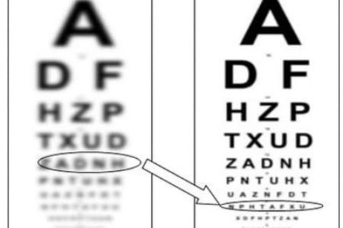
Cataract affects reading sight and the ability to clearly see nearby objects in the same way as in the case of presbyopia. But if a person suffering due to presbyopia caused by lens elasticity loss can strengthen the eyes with specially-created exercises, adding vitamins and adjusting diet, and correcting the poor vision with reading glasses, in the case of cataract, such a healing strategy won't work. Thus, noticing the following alarming signs, schedule an appointment with an eye doctor to avoid complete loss of vision:
- Foggy and dim vision and loss of visual recognition of things.
- Too high sensitivity to light, so you may experience painful sensations when your eye interacts with bright light.
- Colors seem faded, dull, and washed out.
- You've noticed that you cannot see well at night or in poor light meaning that your eye is unable to adapt to poorly illuminated surroundings.
- There is a need for brighter lighting when reading.
- The blurred image is not fixed even when wearing corrective glasses.
- Double vision is possible.
Not all signs are listed above. It is of paramount importance to understand that the same eye changes may indicate the development of other dangerous diseases.
Even having no complaints, every responsible person that falls into the 40 years old category must undergo regular examination by an ophthalmologist. You need to be examined once every 2-3 years. If pathology is detected, a further regular check-up is imperative. The earlier a disease will be diagnosed, the smoother the treatment will be and the more favorable the prognosis.
Cataract causes & risk factors: protect yourself from the harmful outcome
There are lots of techniques on how to improve eyesight which can help you to cope with lots of eye troubles. Unfortunately, lens opaqueness is not included in this group of illnesses. So what are those factors that can provoke this medical issue? Unfavorable environmental conditions, constant stress, the intense rhythm of life, chronic diseases, and metabolic disorders - all these are prior conditions for the development of the disease. There is only one piece of advice: to consult an ophthalmologist as soon as possible, as even if the disorder is considered age-related, eye lens clouding can occur at any age, and the category of predisposing factors are quite extensive:
- inflammatory eye disease;
- hereditary predisposition;
- eye injuries and contusions;
- concomitant eye diseases, such as glaucoma;
- chemical, ultraviolet, or radiation effects on the body;
- endocrine diseases (diabetes mellitus, decreased production of the vitally important hormones, overactivity of the thyroid gland, etc.);
- consequences of severe infections and intoxications;
- bad habits, including smoking and alcoholism;
- immune system dysfunction.
Cataract treatment and the possibility of surgery
Is drug therapy effective?
Eye drops help to improve metabolic processes in the lens and eye tissues, due to which the progressive eye impairment can be slowed down. However, it is crucial to understand that modern medicine does not have a drug that can solve the problem and regain perfect sight without cataract removal.
The most common surgical procedure
With modern surgical techniques, it is possible to operate a patient at any stage, although the minimum standards must be still maintained. Thus, with a slight decrease in vision, the operation usually is not performed. Severe visual acuity worsening, leading to disability and discomfort in everyday life, are the main indicators to correct the vision with surgery.
The operation to replace the lenses with artificial ones takes about 15 minutes, it is performed under local anesthesia, while the patient does not experience any inconveniences. Efficient steps in such surgery can be performed using the impressive capabilities of the femtosecond laser. An unprecedented high accuracy, the gentlest, delicate effect on the structures of the eye, the short recovery from cataract surgery are just some of the advantages.
Tips for the post-surgery period
There are general recommendations, but it is vital to find out the best steps from the eye doctor to minimize the time of recovery through the individual plan, taking into account how the operation went:
- use the aftercare eye drops as prescribed;
- monitor your condition (remember to clarify which symptoms are normal and don’t require further treatment, and which are abnormal and must be handled properly);
- do not sleep on the side of the operated eye during the first days;
- avoid swimming and temperature fluctuations;
- do not lift heavy things and don't do strenuous exercises for at least three months (specify a more precise period in your doctor);
- in case of extreme and intense sensitivity to light, use sunglasses;
- patients are allowed to watch TV and read, however, they must be ready that objects may be not clearly or distinctly visible.
After surgery, the body needs time to get used to the artificial lens.
The disease can affect one or both eyes. In the second case, it can develop symmetrically, but in one eye it will be more pronounced. If you struggle with cataracts in both eyes, the procedure is performed on one eye first. After a break between operations for at least two weeks, the second eye can be also treated surgically. This is due to the fact that sight in one eye should fully stabilize before the next step.
Are there chances to prevent the disease?
If clouding of the image appears in the lens, then, alas, the process is impossible to reverse. However, in order to prevent illness occurrence, the above-mentioned risk factors, such as diabetes mellitus, dermatitis, eye injuries (contusions), chronic inflammation of the retina, poor lifestyle choices, such as tobacco consumption, must be also competently managed to mitigate all risks. To prevent the formation of various types of cataracts, living healthy and annual observation by an ophthalmologist are key points.