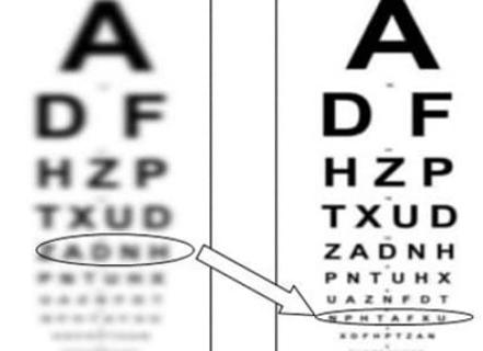
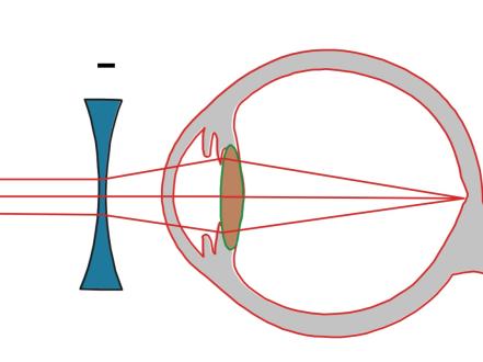
Myopia (nearsightedness) is a refractive error in which parallel light focuses in front of the retina, causing blurred distance vision with clear near vision. It typically results from increased axial length of the eye and develops in childhood, often progressing through adolescence. Diagnosis is by refraction (ideally cycloplegic in children) and ocular examination. Treatment is optical correction (spectacles or contact lenses), with additional options to slow progression in children (eg, low-dose atropine [off-label], dual-focus soft contact lenses, orthokeratology). High/pathologic myopia can lead to retinal and macular complications.
Key Points
- Myopia is caused most commonly by excessive axial elongation of the eye; distance vision is blurred and near vision is relatively clear.
- Cycloplegic refraction is required to accurately diagnose and stage myopia in children and to unmask pseudomyopia.
- Pediatric myopia progression can be moderated by evidence-based measures (eg, low-dose atropine [off-label in the US], dual-focus daily disposable soft contact lenses [FDA-approved], orthokeratology [off-label], and behavioral strategies including increased outdoor time).
- High myopia (≥ −6.00 D or axial length ≥ 26.0–26.5 mm) carries increased lifetime risks of retinal detachment, myopic maculopathy/choroidal neovascularization, glaucoma, and early cataract.
- Red flags include new flashes/floaters or a curtain of vision loss (possible retinal tear/detachment) and severe eye pain with halos and blurry vision (possible acute angle closure from medication-induced ciliary body effusion).
Anatomy and Physiology
The eye’s optical power derives from the cornea (approximately two-thirds) and crystalline lens (approximately one-third). In emmetropia, the optical power and the axial length (~24 mm in adults) align such that distant objects focus on the fovea. In myopia, excessive total refractive power or, most commonly, increased axial length shifts the focal point anterior to the retina, degrading distance acuity. Axial elongation can thin the choroid and sclera and stretch the posterior pole.
Etiology
- Axial elongation (predominant mechanism)
- Corneal steepening (eg, keratoconus)
- Lens-induced changes (eg, nuclear sclerosis, osmotic lens swelling in hyperglycemia)
- Medication-related ciliary body/choroidal effusion with forward lens-iris diaphragm shift (eg, topiramate), causing acute myopic shift
- Genetic predisposition (family history, polygenic)
- Environmental/behavioral factors: reduced outdoor time, intensive near work, urbanization
Pathophysiology
Myopia onset and progression reflect a mismatch between ocular growth and emmetropization. Visual signals at the peripheral retina are hypothesized to drive axial elongation. Scleral remodeling (reduced collagen cross-linking, altered extracellular matrix) and choroidal thinning accompany progressive myopia. High/pathologic myopia leads to posterior staphyloma, lacquer cracks (Bruch’s membrane breaks), myopic choroidal neovascularization (mCNV), and increased susceptibility to peripheral retinal degenerations (lattice, atrophic holes).
Epidemiology
- In US surveys, the prevalence of myopia in people 12 to 54 years increased from 25.0% (1971–1972) to 41.6% (1999–2004) [1].
- Globally, myopia is projected to affect ~50% of the world’s population by 2050, with ~10% having high myopia [2].
- Onset most often occurs between ages 6–12 years; progression typically decelerates in late adolescence/early adulthood.
- Risk factors: parental myopia, East Asian ancestry, limited outdoor time, high educational intensity, prematurity/retinopathy of prematurity.
Classification
- By degree
- Low: < −3.00 diopters (D)
- Moderate: −3.00 to −6.00 D
- High: ≥ −6.00 D (often correlates with axial length ≥ 26.0–26.5 mm)
- By age of onset
- Early-onset (preschool/early school-age)
- School-age (most common)
- Adult-onset (eg, occupational near work–related)
- By pathologic features
- Simple (no structural complications)
- Pathologic/degenerative (posterior staphyloma, myopic maculopathy, mCNV)
- By refractive component
- Axial, corneal (eg, keratoconus), lenticular (eg, nuclear sclerosis)
Symptoms and Signs
Simple/axial myopia
- Symptoms: blurred distance vision, squinting, eye strain (asthenopia), headaches with prolonged visual tasks.
- Pediatric cues: sitting close to screens/board, difficulty in distance-dependent activities (sports, driving simulators in teens), eye rubbing.
- Signs: decreased unaided distance acuity; improved acuity with pinhole; normal anterior segment; fundus tessellation, tilted disc, peripapillary atrophy may be present.
High/pathologic myopia
- Symptoms: metamorphopsia, paracentral scotomas, new floaters/flashes, peripheral field defects.
- Signs: posterior staphyloma, lacquer cracks, myopic macular atrophy, Fuchs spots, chorioretinal atrophy; peripheral lattice degeneration, atrophic holes; posterior vitreous detachment at younger age.
Secondary/myopic shift
- Acute myopic shift: sudden blur at distance and near, halos if angle becomes shallow; often medication-triggered (eg, topiramate) or due to lens changes (hyperglycemia, early nuclear sclerosis).
Complications
- Retinal tears and rhegmatogenous retinal detachment (risk increases with higher myopia and longer axial length) [3].
- Myopic maculopathy, including patchy atrophy and mCNV, causing central vision loss [4].
- Open-angle glaucoma association and earlier posterior vitreous detachment [5].
- Early nuclear sclerotic cataract and lens instability in extreme axial elongation.
- Contact lens–related infectious keratitis (particularly with overnight wear/orthokeratology) [6].
Diagnosis
Clinical evaluation
- History: age of onset, progression rate, family history, near work/outdoor time, prior treatments (atropine, orthokeratology), contact lens habits, medications (eg, topiramate, sulfonamides), systemic disease (diabetes), trauma.
- Visual acuity with and without pinhole; cover tests for phoria/tropia if symptomatic.
- Refraction
- Objective: autorefraction and retinoscopy.
- Subjective refinement.
- Cycloplegic refraction in children/teens and suspected accommodative spasm: cyclopentolate 1% (1 drop OU, repeat in 5 minutes; refraction 30–45 minutes later).
- Slit-lamp exam for anterior segment, lens status.
- Dilated fundus examination for peripheral lesions, macular changes, and optic nerve.
Imaging and laboratory testing
- Axial length and keratometry via optical biometry (eg, partial coherence interferometry) to monitor progression and surgical planning.
- Optical coherence tomography (OCT) for myopic maculopathy, mCNV, and posterior staphyloma assessment.
- Widefield fundus photography for documentation of peripheral lesions.
- B-scan ultrasonography if media opacity or to delineate posterior staphyloma.
- Laboratory testing is not routinely indicated unless secondary causes suspected (eg, uncontrolled diabetes).
Differential diagnosis
| Entity | Distinguishing features |
|---|---|
| Pseudomyopia (accommodative spasm) | Variable refraction; near work–induced blur; resolves with cycloplegia; may have esophoria. |
| Keratoconus/ectasia | Irregular astigmatism; scissoring reflex on retinoscopy; corneal topography abnormal; progressive. |
| Nuclear sclerotic cataract | Adult/older patient; myopic shift (“second sight”); lens changes on slit-lamp. |
| Diabetic lens-induced myopia | Fluctuating refraction with hyperglycemia; improves with glycemic control. |
| Medication-induced myopic shift (eg, topiramate) | Acute onset with possible angle narrowing, elevated IOP; resolves after drug cessation. |
| Uncorrected hyperopia with accommodative spasm | Young patients; headaches; cycloplegic refraction reveals latent hyperopia. |
Treatment
Medical management
- Optical correction
- Spectacles: full correction for distance; consider polycarbonate/high-index lenses in moderate-to-high myopia; ultraviolet protection.
- Contact lenses: daily disposables reduce infection risk; caution with overnight wear due to keratitis risk; counsel on hygiene to reduce bacterial conjunctivitis and keratitis risk (see keratitis and bacterial conjunctivitis pages).
- Myopia control in children (evidence-based)
- Low-dose atropine (off-label, compounded in the US): 0.01–0.05% 1 drop OU qHS (once nightly) for 2–3 years; 0.05% shows greater effect with more side effects; counsel on photophobia and mild near blur; consider photochromic/progressive add spectacles if symptomatic; taper to mitigate rebound [7,8]. Off-label status in the US should be discussed with caregivers.
- Dual-focus daily disposable soft contact lenses (eg, MiSight 1 day): FDA-approved for slowing myopia progression in children; worn daily; avoid overnight wear; reinforce hygiene to mitigate infectious risk [9].
- Orthokeratology (overnight corneal reshaping lenses): improves unaided daytime acuity and can reduce axial elongation; off-label for myopia control in the US; increased risk of microbial keratitis, particularly with poor hygiene [6,10].
- Multifocal soft contact lenses (center-distance designs): off-label for myopia control; randomized trials show reduced progression vs single-vision lenses [7].
- Lifestyle/behavioral measures
- Increase outdoor time (eg, ~2 hours/day when feasible) to reduce risk of onset and possibly slow early progression [11].
- Manage continuous near work (eg, breaks with the 20-20-20 rule; maintain working distance ≥ 12–16 inches).
- Treatment of complications
- Myopic CNV: intravitreal anti-VEGF agents (eg, ranibizumab, aflibercept) are first-line; dosing is individualized (usually PRN after loading), with favorable anatomical/visual outcomes [12].
- Retinal tears/detachment: prompt referral for laser retinopexy or retinal surgery (pneumatic retinopexy, scleral buckle, pars plana vitrectomy) per standard indications.
Notes:
- Avoid initiating or continue only with caution medications known to cause acute myopic shift/angle closure (eg, topiramate) in symptomatic patients; urgent evaluation if severe pain/blur/halos.
Procedural and surgical management
- Refractive surgery (adults with stable refraction)
- Laser vision correction: LASIK, PRK, SMILE for suitable corneal/ocular anatomy; counseled risks include dry eye, ectasia, glare/halos; not performed in progressing pediatric myopia.
- Phakic intraocular lens (ICL) implantation for high myopia with adequate anterior chamber depth and endothelial cell counts.
- Clear lens extraction is rarely used and sacrifices accommodation; reserved for selected cases with counseling on retinal detachment risk.
- Myopia control claims are not established for refractive surgery.
- Coding varies by payer and technique.
- Pathologic myopia interventions
- Posterior staphyloma and macular tractional changes: consider vitrectomy with/without macular buckling in selected cases by a retina specialist.
Special populations
- Pediatric: Use cycloplegic refraction for accuracy; prioritize evidence-based myopia control where appropriate; shared decision-making with caregivers regarding off-label atropine and contact lens options; close follow-up (eg, every 6 months with refraction and axial length when available).
- Pregnancy/lactation: Corneal sensitivity and hydration change; defer elective refractive surgery until several months postpartum; safety data for chronic low-dose atropine in pregnancy/lactation are limited—avoid unless potential benefit justifies potential risk; consider nonpharmacologic options [FDA labeling consultation recommended].
- High myopia/elderly: Vigilant monitoring for maculopathy, glaucoma, and cataract; lower threshold for retina referral if new symptoms.
Prognosis
Most simple myopia stabilizes by early adulthood with excellent corrected vision. High/pathologic myopia confers cumulative risks of sight-threatening complications that require lifelong surveillance. In children, optical and pharmacologic myopia control strategies reduce average progression and axial elongation versus single-vision correction, though individual responses vary and treatment effects diminish after cessation with possible rebound [7,8,10].
Prevention and Patient Counseling
- Encourage regular comprehensive eye examinations (cycloplegic refraction in children).
- Promote outdoor time and healthy near-work habits in school-age children.
- Discuss evidence-based myopia control options, on-label versus off-label status, benefits, risks, and adherence.
- Reinforce contact lens hygiene; avoid overnight wear unless specifically indicated and supervised (orthokeratology).
- Educate high myopes on symptoms of retinal tear/detachment and the need for urgent evaluation.
When to Seek Immediate Care
- Sudden showers of floaters, flashes of light, or a curtain/shadow in your side vision.
- Sudden, severe eye pain with headache, halos around lights, nausea/vomiting, and blurry vision.
- A sudden drop in vision in one or both eyes.
- Painful red eye while wearing contact lenses, especially with light sensitivity.
- Recent eye trauma with new visual symptoms.
References
- Vitale S, Sperduto RD, Ferris FL III. Increased prevalence of myopia in the United States between 1971–1972 and 1999–2004. Arch Ophthalmol. 2009;127(12):1632-1639. doi:10.1001/archophthalmol.2009.303.
- Holden BA, Fricke TR, Wilson DA, et al. Global prevalence of myopia and high myopia and temporal trends from 2000 through 2050. Ophthalmology. 2016;123(5):1036-1042. doi:10.1016/j.ophtha.2016.01.006.
- Haarman AEG, Enthoven CA, Tideman JWL, et al. The complications of myopia: a review and meta-analysis. Invest Ophthalmol Vis Sci. 2020;61(4):49. doi:10.1167/iovs.61.4.49.
- Ohno-Matsui K, Wu PC, Yamashiro K, et al. IMI Pathologic Myopia. Invest Ophthalmol Vis Sci. 2021;62(5):PMID 33978354. doi:10.1167/iovs.62.5.5.
- Marcus MW, de Vries MM, Montolio FG, Jansonius NM. Myopia as a risk factor for open-angle glaucoma: a systematic review and meta-analysis. Ophthalmology. 2011;118(10):1989-1994. doi:10.1016/j.ophtha.2011.03.012.
- Stapleton F, Keay L, Edwards K, et al. The incidence of contact lens-related microbial keratitis in Australia. Ophthalmology. 2008;115(10):1655-1662. doi:10.1016/j.ophtha.2008.04.002. [Representative of increased risk with overnight wear]
- AAO Clinical Statement. Myopia Control in Children. American Academy of Ophthalmology; 2022. https://www.aao.org/clinical-statement/myopia-control-in-children
- Yam JC, Li FF, Zhang X, et al. Two-year clinical trial of the low-concentration atropine for myopia progression (LAMP) study. Ophthalmology. 2020;127(7):910-919. doi:10.1016/j.ophtha.2019.12.011.
- U.S. FDA. FDA approves first contact lens indicated to slow the progression of nearsightedness in children. 2019. https://www.fda.gov/news-events/press-announcements/fda-approves-first-contact-lens-indicated-slow-progression-nearsightedness-children
- Huang J, Wen D, Wang Q, et al. Efficacy comparison of 16 interventions for myopia control in children: a network meta-analysis. Ophthalmology. 2016;123(4):697-708. doi:10.1016/j.ophtha.2015.11.010.
- He M, Xiang F, Zeng Y, et al. Effect of time spent outdoors at school on the development of myopia among children in China: a randomized clinical trial. JAMA. 2015;314(11):1142-1148. doi:10.1001/jama.2015.10803.
- Ohno-Matsui K, Yannuzzi LA, Spaide RF. Myopic choroidal neovascularization: diagnosis and treatment. Retina. 2015;35(8):1612-1621. doi:10.1097/IAE.0000000000000602.
ICD-10-CM Codes:
- H52.10 Myopia, unspecified eye
- H52.11 Myopia, right eye
- H52.12 Myopia, left eye
- H52.13 Myopia, bilateral
Disclaimer: This article is for informational purposes and is not a substitute for professional medical advice, diagnosis, or treatment. Always consult a qualified eye care professional about your specific condition. If you have sudden vision loss, severe eye pain, or an eye injury, seek emergency care immediately.