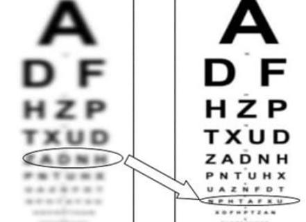
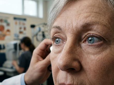
Nystagmus is an involuntary, rhythmic oscillation of the eyes that can be horizontal, vertical, torsional, or mixed. It may be present from infancy (infantile/early-onset) or acquired later due to neurologic, vestibular, visual, metabolic, or drug-related causes. Patients may have blurred vision, reduced visual acuity, head turns (to use a null point where nystagmus dampens), and, in acquired cases, oscillopsia (illusory movement of the environment). Diagnosis is clinical, guided by a detailed ocular and neurologic examination and targeted testing. Management addresses the underlying cause and may include optical strategies, prism, pharmacologic therapy (mostly off-label), and strabismus surgery to reduce abnormal head posture and improve function.
Key Points
- Nystagmus is a sign, not a diagnosis; etiologies span infantile ocular motor disorders, sensory visual deprivation, vestibular disease, central nervous system lesions, medications, and toxins.
- Infantile nystagmus syndrome (INS) typically lacks oscillopsia and often improves with convergence; acquired nystagmus commonly causes oscillopsia and warrants prompt evaluation for neurologic or vestibular disease.
- Clinical characterization (direction, waveform, Alexander’s law, effect of gaze, fixation, convergence) narrows the differential and guides testing.
- Treat reversible causes (eg, drug toxicity, thiamine deficiency, acute vestibular neuritis). Visual rehabilitation, prisms, and selective surgeries improve function in INS.
- Pharmacologic therapy is condition-specific and largely off-label (eg, baclofen for periodic alternating nystagmus; 4-aminopyridine for downbeat nystagmus; gabapentin or memantine in some infantile/acquired cases).
- Sudden-onset nystagmus with brainstem/cerebellar signs, severe vertigo, new diplopia, or headache is an emergency; evaluate for stroke or posterior fossa pathology.
Anatomy and Physiology
Gaze stability depends on integrated ocular motor systems:
- Vestibulo-ocular reflex (VOR): stabilizes images during head motion.
- Neural integrator (brainstem/cerebellar network): holds eccentric gaze; failure yields gaze-evoked nystagmus.
- Smooth pursuit and saccadic systems: track moving targets and generate rapid refixations.
- Optokinetic system: sustains tracking for large-field motion.
- Foveation periods: brief intervals of minimal retinal slip allow best acuity; altered foveation degrades vision in nystagmus.
Cerebellar nodulus/uvula, flocculus, and brainstem nuclei (vestibular, interstitial nucleus of Cajal) are key modulators; lesions produce characteristic nystagmus patterns.
Etiology
-
Infantile/early-onset
- Infantile nystagmus syndrome (INS; idiopathic/motor).
- Sensory nystagmus due to reduced afferent input: albinism, foveal hypoplasia, achromatopsia, Leber congenital amaurosis, aniridia, congenital cataract/corneal opacity, optic nerve hypoplasia or atrophy.
- Latent/manifest latent nystagmus with infantile strabismus (often esotropia).
- Spasmus nutans (triad: nystagmus, head nodding, torticollis) in infancy; typically self-limited.
-
Acquired
- Peripheral vestibular disorders: vestibular neuritis, Ménière disease, labyrinthitis, vestibular schwannoma.
- Central lesions: stroke (especially posterior circulation), demyelination (multiple sclerosis), Chiari malformation, Wernicke encephalopathy, cerebellar degeneration, tumors (brainstem, cerebellum, parasellar/chiasmal), trauma.
- Drug/toxin: anticonvulsants (phenytoin, carbamazepine), lithium, sedatives/hypnotics, alcohol, aminopyridines (excess), toluene.
- Miscellaneous: oculopalatal tremor (hypertrophic olivary degeneration), thyroid eye disease (rare pendular), superior oblique myokymia (not true nystagmus).
Pathophysiology
- Jerk nystagmus: slow drift due to imbalance (eg, vestibular tone asymmetry or leaky neural integrator) followed by a fast corrective saccade. Alexander’s law: intensity increases in the direction of the fast phase (typical for peripheral vestibular lesions).
- Pendular nystagmus: sinusoidal, no fast phase; arises from instability of oscillatory neural networks (eg, acquired pendular nystagmus in multiple sclerosis or oculopalatal tremor; pendular waveforms also common in INS).
- Waveform attributes (amplitude, frequency, foveation quality) correlate with visual acuity in INS; convergence often dampens INS via vergence-related mechanisms.
- Specific patterns localize lesions: downbeat (craniocervical junction/cerebellar flocculus), upbeat (brainstem), periodic alternating (cerebellar nodulus/uvula), seesaw (parasellar/chiasmal), convergence-retraction “nystagmus” (dorsal midbrain—actually a saccadic disorder).
Epidemiology
- INS prevalence is estimated at ~1 in 1,000 to 1 in 1,500 people in Western populations.
- Afferent visual pathway disorders (eg, albinism) account for a substantial proportion of INS in pediatric cohorts; idiopathic/motor INS is also common [1,2].
- Acquired nystagmus incidence is not well defined; it is frequent in neuro-otology clinics and in multiple sclerosis.
Classification
- By age of onset: infantile (≤6 months) vs acquired (>6 months).
- By waveform: jerk vs pendular.
- By primary plane: horizontal, vertical (downbeat, upbeat), torsional, mixed.
- By behavior:
- Gaze-evoked vs gaze-independent.
- Fixation-suppressed (peripheral vestibular) vs not suppressed (central).
- Periodic alternating (cycles direction every ~90–120 minutes).
- Latent/manifest latent (emerges or increases with monocular occlusion).
- Convergence-dampened vs convergence-enhanced.
- By association: with strabismus, head posture, afferent defects.
Symptoms and Signs
-
Infantile nystagmus syndrome (INS)
- Onset in early infancy; horizontal predominant; pendular or jerk with accelerating slow phases.
- Often improves with convergence and in near fixation; absent oscillopsia.
- Null zone with abnormal head posture (face turn, chin up/down).
- Variable visual acuity; associated sensory diagnoses (eg, albinism) reduce acuity further.
- May have manifest latent component if strabismus is present.
-
Sensory nystagmus (infantile)
- Early onset with decreased visual behavior; fundus, OCT, ERG, or genetic testing often reveal the underlying cause (eg, foveal hypoplasia).
-
Spasmus nutans
- Asymmetric high-frequency, low-amplitude nystagmus with head nodding and torticollis; typically resolves by age 3–4; atypical features warrant imaging.
-
Peripheral vestibular nystagmus
- Acute onset, horizontal-torsional jerk nystagmus; follows Alexander’s law; suppresses with fixation; associated vertigo, nausea, gait unsteadiness; direction fixed relative to head.
-
Central nystagmus
- Pure vertical (downbeat/upbeat) or pure torsional; direction-changing gaze-evoked nystagmus; not suppressed by fixation; often with other neurologic signs (dysarthria, limb ataxia, diplopia).
-
Acquired pendular nystagmus
- Often binocular, multidirectional; disabling oscillopsia; associated with multiple sclerosis or oculopalatal tremor.
-
Localizing patterns
- Downbeat: craniocervical junction/cerebellar disease.
- Upbeat: medullary/paramedian brainstem lesions.
- Periodic alternating: cerebellar nodulus/uvula dysfunction or idiopathic.
- Seesaw: parasellar/chiasmal region.
- Convergence-retraction: dorsal midbrain (Parinaud) syndrome.
Complications
- Reduced visual acuity and contrast sensitivity; impaired reading speed and visuomotor function.
- Abnormal head posture leading to neck strain and musculoskeletal pain.
- Psychosocial impact and functional limitations (eg, driving eligibility varies by jurisdiction).
- Risk of amblyopia in children with associated sensory disorders or strabismus.
- Falls and disability in patients with severe oscillopsia.
Diagnosis
Clinical evaluation
- History: age at onset; variability with gaze, fixation, convergence, near vs distance; oscillopsia; vertigo; diplopia; neurologic symptoms; developmental history; family history; medications/toxins; birth/trauma history.
- Examination:
- Characterize waveform, direction, and intensity in primary position and eccentric gazes; apply fixation (Frenzel goggles if needed) and observe Alexander’s law.
- Assess suppression with fixation and effect of convergence (near target; base-out prism test).
- Look for null point and abnormal head posture.
- Ocular exam: visual acuity (preferably with nystagmus acuity function testing when available), refraction (cycloplegic in children), ocular alignment/versions, slit-lamp, intraocular pressure, dilated fundus.
- Neurologic screen: cranial nerves, cerebellar testing, gait, sensory/motor exam.
Imaging and laboratory testing
- Eye movement recording: video-oculography/electronystagmography to quantify waveform and response to maneuvers [1].
- Optical coherence tomography (OCT): foveal hypoplasia, optic nerve anatomy; helpful in albinism and aniridia [2].
- Visual electrophysiology: electroretinography (ERG) for retinal dystrophies; visual evoked potentials (VEP) for optic pathway dysfunction [2].
- Genetic testing when features suggest syndromic or inherited disease (eg, FRMD7 in X-linked INS; genes for albinism/achromatopsia) [2].
- Neuroimaging (MRI brain ± orbits with contrast):
- Mandatory in acquired nystagmus, central/localizing patterns (downbeat, upbeat, seesaw), neurologic signs, or atypical/late-onset “spasmus nutans–like” presentations to exclude optic pathway glioma [1,3].
- Vestibular testing (video head impulse, calorics) for suspected peripheral vestibular etiologies.
- Targeted labs when indicated (eg, thiamine deficiency, lithium level, autoimmune/metabolic panels).
Differential diagnosis
| Entity | Distinguishing features |
|---|---|
| Physiologic end-gaze nystagmus | Low amplitude at extreme gaze; absent in primary; no symptoms. |
| Saccadic intrusions (square-wave jerks) | Brief saccades away and back without slow phase; common in cerebellar disease and the elderly. |
| Opsoclonus/ocular flutter | Chaotic or back-to-back saccades without intersaccadic interval; no slow phase; paraneoplastic or post-viral. |
| Superior oblique myokymia | Monocular oscillopsia; fine high-frequency torsional-vertical tremor; intermittent. |
| Voluntary nystagmus | Rapid, low-amplitude oscillations on command; fatigues; normal exam otherwise. |
| Convergence-retraction “nystagmus” | Jerky convergence with retraction on upgaze attempts; dorsal midbrain lesion; not true nystagmus. |
| Bilateral vestibulopathy | Oscillopsia with head motion; abnormal VOR; no spontaneous nystagmus at rest. |
| Ocular myokymia/eyelid myokymia | Fasciculations of extraocular or eyelid muscles; does not move the globe rhythmically. |
Treatment
Medical management
-
Correct underlying cause
- Discontinue or reduce offending drugs/toxins (eg, anticonvulsants, lithium, sedatives) when feasible [4].
- Thiamine for Wernicke encephalopathy; disease-specific therapy for demyelination, stroke, or tumor.
- Vestibular neuritis: vestibular rehabilitation; short-term vestibular suppressants may palliate vertigo but can impede central compensation if prolonged [5].
-
Optical/rehabilitative interventions
- Full refractive correction; consider contact lenses (may improve vision and dampen nystagmus in some INS due to enhanced proprioceptive feedback) [2].
- Filters/tints for photosensitivity in albinism; low-vision services as needed.
- Prisms:
- Yoked prisms to shift the image into the null zone and reduce abnormal head posture.
- Base-out prisms to induce convergence in INS patients with convergence damping [2].
-
Pharmacotherapy (largely off-label in the US; discuss risks/benefits and monitor)
- Baclofen 5–10 mg PO tid (three times daily) for periodic alternating nystagmus; may abolish cycles in responders [1,6].
- 4-aminopyridine (dalfampridine) 10 mg PO q12h for downbeat nystagmus; improves slow-phase velocity and symptoms in small trials; avoid in seizure history or severe renal impairment (FDA-labeled indication is gait in MS; use here is off-label) [7,8].
- Clonazepam 0.25–0.5 mg PO bid for downbeat or upbeat nystagmus when aminopyridines are not tolerated [1,6].
- Gabapentin 300–600 mg PO tid to qid (900–2,400 mg/day) and/or memantine 10 mg PO bid have shown modest improvements in nystagmus intensity and visual function in some infantile and acquired forms; evidence is mixed; both are off-label [1,6,9].
- Botulinum toxin (retrobulbar or extraocular muscle injections) can transiently reduce oscillations and oscillopsia but often causes ptosis and diplopia; reserve for severe, refractory cases [1,2].
Note: Dosing must be individualized; start low and titrate while monitoring for CNS depression, imbalance, and drug interactions.
Procedural and surgical management
-
Strabismus/nystagmus surgery (primarily for INS)
- Kestenbaum–Anderson procedures (augmented horizontal rectus recessions/resections) to shift the null point toward primary gaze and reduce abnormal head posture; visual acuity may improve modestly through better foveation [2,6].
- Large symmetric recessions or tenotomy and reattachment (TAR) procedures are used in selected cases to reduce nystagmus intensity [2,6].
- Concomitant strabismus correction can improve alignment and binocularity.
- Patient selection: stable, well-defined null point; significant abnormal head posture; realistic expectations regarding acuity gains.
-
Neurosurgical/structural interventions
- Address causative lesions (eg, Chiari I decompression for symptomatic downbeat nystagmus due to cerebellar tonsillar ectopia) [1,6].
Special populations
-
Pediatric
- Early referral (ideally by 2–3 months of age if nystagmus is observed) for comprehensive sensory evaluation, cycloplegic refraction, and imaging/genetic testing when indicated [2,3].
- Treat amblyopia and refractive errors aggressively; consider prisms for convergence damping.
- Spasmus nutans: observe if classic and improving; obtain MRI if onset after 1 year, asymmetric/persistent beyond age 3–4, optic disc pallor, or other atypia to exclude optic pathway glioma [3].
-
Pregnancy and lactation
- Limited safety data for gabapentin, memantine, baclofen, and aminopyridines; weigh maternal benefit against fetal/infant risk; prefer nonpharmacologic measures when possible; coordinate with obstetrics [FDA labels and tertiary sources].
-
Older adults and medically complex patients
- Heightened sensitivity to CNS depressants; increased fall risk with sedating agents; simplify regimens and emphasize vestibular/visual rehabilitation.
Prognosis
- INS is typically lifelong but often stable; many patients achieve functional vision with optical correction, accommodations, and, when appropriate, surgery. Visual acuity depends on waveform/foveation and any associated sensory pathology [2,6].
- Acquired nystagmus outcomes depend on etiology. Vestibular causes often improve as compensation develops. Central or degenerative causes may persist; symptom control is variable but possible with targeted therapy and rehabilitation [1,6].
Prevention and Patient Counseling
- Avoid or minimize sedatives, excess alcohol, and medications known to induce nystagmus when alternatives exist.
- Maintain adequate nutrition; promptly treat conditions such as Wernicke encephalopathy.
- Use optimal refractive correction; consider contact lenses or prisms if recommended.
- Coordinate low-vision services, school/work accommodations, and driving evaluations in accordance with state regulations.
- For caregivers: report early ocular motor abnormalities in infants; timely evaluation identifies treatable sensory causes and prevents amblyopia.
When to Seek Immediate Care
- Sudden-onset jerking eye movements with severe vertigo, new double vision, slurred speech, weakness, numbness, or trouble walking.
- New nystagmus after head trauma.
- New oscillopsia or nystagmus with severe headache, neck pain, or altered mental status.
- Any new nystagmus in an adult without a prior history.
- Nystagmus in an infant noted in the first months of life.
Call 911 or go to the emergency department for the first three situations.
References
- Leigh RJ, Zee DS. The Neurology of Eye Movements, 5th ed. Oxford University Press; 2015.
- EyeWiki. Infantile Nystagmus. American Academy of Ophthalmology. https://eyewiki.aao.org/Infantile_Nystagmus. Accessed July 24, 2026.
- AAPOS. Nystagmus. American Association for Pediatric Ophthalmology and Strabismus. https://aapos.org/glossary/nystagmus. Accessed July 24, 2026.
- StatPearls. Nystagmus. Treasure Island (FL): StatPearls Publishing; updated 2025. https://www.ncbi.nlm.nih.gov/books/NBK470211/
- Fife TD, Iverson DJ, Lempert T, et al. Practice parameter: therapies for benign paroxysmal positional vertigo (an evidence-based review). Neurology. 2008;70(22):2067-2074. doi:10.1212/01.wnl.0000313378.77444.ac. [Contextual vestibular management]
- EyeWiki. Downbeat Nystagmus; Periodic Alternating Nystagmus; Nystagmus Overview. American Academy of Ophthalmology. https://eyewiki.aao.org/Downbeat_Nystagmus; https://eyewiki.aao.org/Periodic_Alternating_Nystagmus; https://eyewiki.aao.org/Nystagmus. Accessed July 24, 2026.
- FDA DailyMed. Dalfampridine (Ampyra) label. https://dailymed.nlm.nih.gov/dailymed/drugInfo.cfm?setid=0b7bde83-2e06-4b50-9b0b-3b8c60a1b11d. Accessed July 24, 2026.
- Kalla R, Muroi A, Schmitt N, et al. 4-Aminopyridine for the treatment of downbeat nystagmus: a randomised, double-masked, placebo-controlled trial. J Neurol Neurosurg Psychiatry. 2011;82(11):1254-1257. doi:10.1136/jnnp.2010.235739.
- Thurtell MJ, Joshi AC, Leone AC, et al. Crossover trial of gabapentin and memantine as treatment for acquired nystagmus. Neurology. 2010;74(11): 1–8.
- Maconachie G, Gottlob I. Nystagmus: state of the art. Clin Exp Optom. 2019;102(6):497-503. doi:10.1111/cxo.12922.
This article is for informational purposes and is not a substitute for professional medical advice, diagnosis, or treatment. Always consult a qualified eye care professional about your specific condition. If you have sudden vision loss, severe eye pain, or an eye injury, seek emergency care immediately.