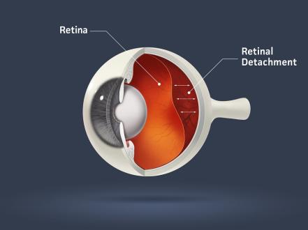
Retinopathy of prematurity (ROP) is a vasoproliferative retinal disorder that affects premature infants with incompletely vascularized retinas. Pathologic neovascularization can lead to tractional retinal detachment and permanent vision loss. ROP is asymptomatic; diagnosis relies on standardized dilated retinal examination in the neonatal intensive care unit (NICU) and after discharge. Management is determined by zone, stage, and plus disease and includes laser photocoagulation or intravitreal anti–vascular endothelial growth factor (VEGF) therapy; advanced stages may require vitreoretinal surgery.
Key Points
- ROP arises from abnormal retinal vessel development in premature infants; the most important risk factors are low gestational age and low birth weight [1,2].
- Screening is guideline-driven: infants ≤30 weeks’ gestation or ≤1,500 g—and selected unstable infants up to 2,000 g—require serial dilated fundus examinations beginning at 31 weeks postmenstrual age (PMA) or 4 weeks chronologic age, whichever is later [1].
- Treatment is indicated for “Type 1 ROP” (per ETROP criteria) and usually uses near-confluent laser to the avascular retina; anti-VEGF is preferred for zone I, posterior zone II, or aggressive ROP [2,3,4].
- FDA-approved intravitreal agents for ROP include ranibizumab (0.2 mg) and aflibercept (0.4 mg); bevacizumab (0.625 mg) is widely used off-label [5–7].
- After anti-VEGF, recurrence may occur late; infants require extended follow-up until vascularization is complete and at least to 65–70 weeks PMA [1,2,4].
- Long-term risks include high myopia, strabismus, amblyopia, and late retinal detachment; ongoing pediatric ophthalmology follow-up is essential [2,8].
Anatomy and Physiology
The retina’s vascularization starts at the optic nerve head around 16 weeks’ gestation and progresses peripherally to the ora serrata by term (40 weeks). Premature birth interrupts physiologic angiogenesis, leaving an avascular peripheral retina. Retinal oxygen demand rises with maturation of photoreceptors; pathologic responses to hypoxia and growth factor imbalance drive neovascularization.
Etiology
- Prematurity with incomplete retinal vascularization.
- Low gestational age and low birth weight (strongest predictors).
- Oxygen exposure and fluctuations in oxygen saturation.
- Systemic illness: sepsis, necrotizing enterocolitis, bronchopulmonary dysplasia, apnea/bradycardia, anemia/transfusions, poor postnatal weight gain/low insulin-like growth factor 1 (IGF-1) [1,2].
- Postnatal factors: hypercarbia, acidosis, poor nutrition.
Pathophysiology
ROP has two phases:
- Phase I (vasoattenuation): After premature birth, relative hyperoxia and low IGF-1 suppress physiologic vascular growth; peripheral retina remains avascular.
- Phase II (vasoproliferation): As metabolic demand increases, the avascular periphery becomes hypoxic, inducing VEGF and other mediators. Pathologic extraretinal neovascularization at the vascular–avascular junction can progress to fibrovascular proliferation, vitreoretinal traction, and retinal detachment [2].
Aggressive ROP (AROP) features rapidly progressive posterior disease with prominent plus disease and flat neovascularization, requiring urgent treatment.
Epidemiology
In the United States, an estimated 14,000–16,000 premature infants develop ROP annually; about 1,100–1,500 require treatment and 400–600 develop legal blindness despite care [8]. Incidence and severity increase with decreasing gestational age and birth weight [1,2]. Improvements in neonatal care have increased survival of extremely low-birth-weight infants, maintaining a stable at-risk population.
Classification
Use the International Classification of Retinopathy of Prematurity, Third Edition (ICROP3) [2]:
- Zones (location):
- Zone I (posterior pole; radius twice the distance from the optic disc to fovea),
- Zone II (from zone I to nasal ora serrata),
- Zone III (residual temporal crescent).
- Stages (severity):
- Stage 1: Demarcation line,
- Stage 2: Ridge,
- Stage 3: Extraretinal fibrovascular proliferation,
- Stage 4A/4B: Partial retinal detachment without/with foveal involvement,
- Stage 5: Total retinal detachment.
- Vascular abnormality: Plus disease (arteriolar tortuosity and venous dilation of the posterior pole) vs pre-plus.
- Extent: Clock hours.
- Aggressive ROP (AROP): Rapid posterior disease with severe vascular changes.
Treatment thresholds (ETROP) [3]:
- Type 1 ROP (treat): Zone I, any stage with plus; Zone I, stage 3 with or without plus; Zone II, stage 2 or 3 with plus.
- Type 2 ROP (observe closely): Zone I, stage 1 or 2 without plus; Zone II, stage 3 without plus.
Symptoms and Signs
ROP is asymptomatic. Findings are detected by indirect ophthalmoscopy with scleral depression:
- Posterior pole vascular dilation and tortuosity (plus disease).
- Demarcation line/ridge at the junction of vascularized and avascular retina.
- Extraretinal neovascularization, preretinal hemorrhage.
- Fibrovascular proliferation with traction.
- Retinal detachment (partial or total) in advanced stages.
Aggressive ROP:
- Posterior location (zone I/posterior zone II), prominent plus disease, flat neovascularization, rapid progression.
Complications
- Tractional retinal detachment (stages 4–5).
- Macular dragging, ectopia of the fovea.
- High myopia, astigmatism, anisometropia (particularly post-laser).
- Amblyopia, strabismus, nystagmus.
- Cataract, glaucoma (angle closure from anterior segment changes).
- Late reactivation after anti-VEGF with risk of recurrent neovascularization.
- Late-onset retinal tears/detachment in adolescence/adulthood due to peripheral vitreoretinal abnormalities [2].
Diagnosis
Clinical evaluation
- Target population: Infants with birth weight ≤1,500 g or gestational age ≤30 weeks, and selected infants with birth weight 1,500–2,000 g or >30 weeks with an unstable clinical course or high-risk factors [1].
- Timing: First exam at 31 weeks PMA or 4 weeks chronologic age, whichever is later; subsequent exams every 1–3 weeks based on findings until complete vascularization or regression [1].
- Technique: Dilated binocular indirect ophthalmoscopy with scleral depression by an experienced ophthalmologist. Documentation of zone, stage, extent (clock hours), and plus status using ICROP3.
Imaging and laboratory testing
- Wide-field digital retinal imaging (e.g., contact camera) is useful for documentation, telemedicine, and education; it does not replace bedside ophthalmoscopy for treatment decisions in many centers [2].
- Optical coherence tomography (OCT) can reveal macular edema and structural changes but is adjunctive.
- Laboratory testing has no role in routine ROP diagnosis.
Differential diagnosis
| Entity | Distinguishing features |
|---|---|
| Familial exudative vitreoretinopathy (FEVR) | Full-term infants; peripheral avascular retina with neovascularization; positive family history; genetic variants (e.g., Wnt pathway); can mimic ROP in older child. |
| Persistent fetal vasculature (PFV) | Unilateral microphthalmia, retrolental fibrovascular tissue, persistent hyaloid artery; no classic ROP ridge; full-term birth. |
| Norrie disease | X-linked; bilateral leukocoria in males; congenital retinal dysplasia/detachment; systemic hearing/cognitive issues. |
| Incontinentia pigmenti | Female infants; retinal neovascularization/exudation; skin lesions in Blaschko lines; dental/CNS anomalies. |
| Coats disease | Typically older boys; unilateral telangiectasia with exudation; no prematurity history. |
| TORCH/postnatal infectious retinitis | Chorioretinal scars or active retinitis; systemic infection signs; not stage/zone pattern of ROP. |
| Vitreous hemorrhage of birth trauma | Self-limited hemorrhage in term infants; no ROP staging features. |
Treatment
Treatment aims to ablate the avascular retina or suppress VEGF to induce regression, prevent traction, and preserve macular structure.
Indications (Type 1 ROP; AROP; selected progressing stage 3 without plus in posterior zones) [2,3].
Medical management
- Anti-VEGF intravitreal injections suppress pathologic neovascularization and preserve peripheral retina for potential physiologic vascularization. Particularly useful in zone I, posterior zone II, and AROP; facilitates media clarity in tunica vasculosa lentis.
Agents and typical dosing:
- Ranibizumab (Lucentis): 0.2 mg (0.02 mL) intravitreal; FDA-approved for ROP (2022) [5]. Retreatment based on activity; recurrence can occur weeks to months later [4,5].
- Aflibercept (Eylea): 0.4 mg (0.01 mL) intravitreal; FDA-approved for ROP (2023) [6]. Retreatment interval per label if persistent/recurrent disease [6].
- Bevacizumab (Avastin): 0.625 mg (0.025 mL) intravitreal; off-label; effective in BEAT-ROP for zone I disease [4,7].
Considerations:
- Monitor closely for reactivation until complete vascularization, often through at least 65–70 weeks PMA [1,2,4].
- Systemic VEGF suppression has been measured after bevacizumab; potential neurodevelopmental effects remain under study and uncertain [7]. Ranibizumab has shorter systemic half-life [5].
Adjunctive care:
- NICU practices to stabilize oxygen saturation and optimize nutrition are managed by neonatology teams and may reduce ROP severity [1,2, VERIFY: oxygen target specifics].
Procedural and surgical management
-
Peripheral retinal ablation (laser photocoagulation): Near-confluent laser to avascular peripheral retina for Type 1 ROP or when anti-VEGF is contraindicated. Effective in halting progression and reducing traction risk, but associated with higher rates of high myopia and peripheral field loss than anti-VEGF [2,3]. Performed under topical anesthesia with sedation or general anesthesia as per NICU protocols.
-
Vitreoretinal surgery for detachment:
- Stage 4A: Lens-sparing pars plana vitrectomy may reattach the retina with better macular outcomes.
- Stage 4B/5: Pars plana or pars plicata vitrectomy ± lensectomy; scleral buckle may be used in select 4A/4B eyes [2]. Prognosis declines with stage 5.
- Surgery is highly specialized; referral to a pediatric vitreoretinal surgeon is recommended.
-
Rescue/combined approaches: Anti-VEGF followed by deferred laser to persistent avascular retina is used in some centers to limit late recurrence while maintaining peripheral field; protocols vary and are evolving [2,4, VERIFY].
Special populations
- Extremely low birth weight (<1,000 g) and <27 weeks’ gestation: Highest risk; require early, frequent exams; consider anti-VEGF for posterior disease with meticulous long-term follow-up [1,2].
- Multiple gestation and infants with severe systemic illness: Higher incidence and more labile courses; coordination with neonatology is critical.
- Pregnancy/lactation: Not applicable to patients; counsel caregivers about follow-up demands.
- Immunocompromised: Not applicable to typical ROP epidemiology; standard aseptic intravitreal technique applies.
- Older children and survivors: Require ongoing surveillance for refractive errors, amblyopia, strabismus, and late retinal pathology [2,8].
Prognosis
Most ROP regresses spontaneously without treatment. With timely therapy for Type 1 ROP, anatomic success rates are high, especially in zone II disease [3]. Zone I/AROP has higher recurrence risk and worse structural outcomes even with optimal therapy [2,4,5]. After anti-VEGF, recurrences can occur later than after laser, necessitating prolonged follow-up [4,5]. Long-term visual function is influenced by macular status, refractive error, and amblyopia management.
Prevention and Patient Counseling
- Prevention focuses on reducing prematurity and optimizing perinatal care (antenatal steroids, infection control, nutrition) and NICU oxygen management per evidence-based protocols [1,2].
- Explain the screening schedule, potential need for urgent treatment, and the importance of not missing appointments during and after NICU discharge.
- Counsel about long-term pediatric ophthalmology follow-up to address refractive errors, amblyopia, and strabismus, and to monitor for late retinal complications.
When to Seek Immediate Care
- New outward signs after discharge, such as a white or gray pupil, sudden eye crossing or wandering, or new rapid eye movements.
- Red, swollen eyes or pus after an eye procedure or injection.
- Your baby stops visually tracking faces or lights compared with prior behavior.
- You missed a scheduled ROP eye exam—call to reschedule right away.
- Any sudden change that worries you. Call your ophthalmologist or the NICU team; call 911 for severe illness or trauma.
References
-
Fierson WM; American Academy of Pediatrics Section on Ophthalmology; American Academy of Ophthalmology; American Association for Pediatric Ophthalmology and Strabismus. Screening Examination of Premature Infants for Retinopathy of Prematurity. Pediatrics. 2018;142(6):e20183061. doi:10.1542/peds.2018-3061.
-
Chiang MF, Quinn GE, Fielder AR, et al. International Classification of Retinopathy of Prematurity, Third Edition (ICROP3). Ophthalmology. 2021;128(10):e51–e68. doi:10.1016/j.ophtha.2021.05.031.
-
Early Treatment for Retinopathy of Prematurity Cooperative Group. Revised indications for the treatment of retinopathy of prematurity. Arch Ophthalmol. 2003;121(12):1684–1694. doi:10.1001/archopht.121.12.1684.
-
Mintz-Hittner HA, Kennedy KA, Chuang AZ; BEAT-ROP Cooperative Group. Efficacy of intravitreal bevacizumab for stage 3+ retinopathy of prematurity. N Engl J Med. 2011;364(7):603–615. doi:10.1056/NEJMoa1007374.
-
Stahl A, Lepore D, Fielder A, et al. Ranibizumab versus laser therapy for retinopathy of prematurity (RAINBOW): an open-label randomised controlled trial. Lancet. 2019;394(10208):1551–1559. doi:10.1016/S0140-6736(19)31344-3.
-
U.S. Food and Drug Administration. FDA approves Eylea (aflibercept) to treat retinopathy of prematurity in preterm infants. 2023. https://www.fda.gov/news-events/press-announcements.
-
U.S. Food and Drug Administration. FDA approves Lucentis (ranibizumab) for retinopathy of prematurity. 2022. https://www.fda.gov/news-events/press-announcements.
-
National Eye Institute (NEI). Retinopathy of Prematurity. https://www.nei.nih.gov/learn-about-eye-health/eye-conditions-and-diseases/retinopathy-prematurity. Accessed July 24, 2026.
-
Tong AY, El-Dairi MA, Maldonado RS, et al. Optical coherence tomography in retinopathy of prematurity. Surv Ophthalmol. 2021;66(6):955–970. doi:10.1016/j.survophthal.2021.01.007. [Supports imaging adjuncts]
Disclaimer: This article is for informational purposes and is not a substitute for professional medical advice, diagnosis, or treatment. Always consult a qualified eye care professional about your specific condition. If you have sudden vision loss, severe eye pain, or an eye injury, seek emergency care immediately.