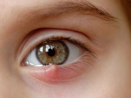
A stye (medical term: hordeolum) is an acute, localized bacterial infection of an eyelid gland, presenting as a tender, red, focal swelling that may point with a pustule. Most styes are caused by Staphylococcus aureus. Diagnosis is clinical. First-line treatment is warm compresses and eyelid hygiene; topical antibiotic ointment may be added for drainage at the lid margin, and incision and drainage is reserved for persistent or complicated cases.

Key Points

  • A stye is an acute bacterial infection of an eyelid gland; external styes involve lash-line glands, internal styes involve meibomian glands.
  • Most cases resolve with warm compresses and eyelid hygiene; do not squeeze or lance at home.
  • Topical antibiotic ointment can be used when there is lash-line drainage; systemic antibiotics are indicated for spreading cellulitis or systemic risk factors.
  • Distinguish from chalazion (painless, firm, chronic nodule) and from preseptal/orbital cellulitis (diffuse swelling, systemic signs).
  • Recurrent styes suggest underlying blepharitis, meibomian gland dysfunction, or rosacea; treat the lid disease to prevent recurrence.
  • Urgent evaluation is needed for fever, worsening diffuse swelling, reduced vision, painful eye movements, or lack of improvement after 48–72 hours of appropriate care.

Anatomy and Physiology

  • Eyelid margin contains:
    • Glands of Zeis (sebaceous) and Moll (apocrine) associated with eyelashes.
    • Meibomian glands embedded in the tarsal plate, secreting meibum that stabilizes the tear film.
  • External hordeolum arises from Zeis or Moll gland infection at the lash line.
  • Internal hordeolum arises from infected meibomian gland within the tarsus, pointing toward the palpebral conjunctiva.

Etiology

  • Usual pathogen: Staphylococcus aureus; methicillin-resistant S. aureus (MRSA) is an occasional cause.
  • Predisposing conditions:
    • Chronic anterior/posterior blepharitis, meibomian gland dysfunction, ocular rosacea, seborrheic dermatitis.
    • Contact lens wear, contaminated cosmetics, poor eyelid hygiene.
    • Diabetes mellitus, immunosuppression.
    • Demodex infestation (Demodex folliculorum) associated blepharitis.

Pathophysiology

  • Obstruction of a lash-line (Zeis/Moll) or meibomian gland orifice allows bacterial overgrowth.
  • Focal suppurative inflammation produces a painful papule/pustule (external) or tender tarsal swelling (internal).
  • Internal hordeola can rupture through conjunctiva or skin or organize into a chronic lipogranulomatous nodule (chalazion).

Epidemiology

  • Styes are common across all ages in primary eye care; exact US incidence and prevalence are not well defined.
  • Recurrent disease correlates with chronic blepharitis and meibomian gland dysfunction.

Classification

  • By site:
    • External hordeolum (stye): Zeis/Moll gland at lash line.
    • Internal hordeolum: meibomian gland in tarsal plate.
  • By course:
    • Acute uncomplicated.
    • Recurrent (≥3/year) often with chronic lid disease.
    • Complicated: associated preseptal cellulitis, abscess, or progression to chalazion.

Symptoms and Signs

  • General:

    • Acute focal eyelid pain, tenderness, erythema, and swelling.
    • Tearing, foreign-body sensation, mild photophobia.
    • Localized yellow pustule (external) or focal tarsal induration (internal).
  • External hordeolum:

    • Tender, erythematous papule at lash follicle with a visible pustular head.
    • Crusting, lash matting, adjacent blepharitis.
  • Internal hordeolum:

    • Diffuse, tender tarsal swelling; may “point” on the palpebral conjunctiva.
    • Conjunctival hyperemia; meibomian gland plugging.
  • Complicated or spreading infection:

    • Preseptal cellulitis: diffuse lid edema, warmth, tenderness without proptosis or ophthalmoplegia.
    • Orbital cellulitis (rare from a stye): fever, proptosis, painful or limited extraocular movements, decreased vision.

Complications

  • Chalazion (chronic lipogranuloma) after resolution of acute infection.
  • Preseptal cellulitis; rarely orbital cellulitis.
  • Abscess formation, lash follicle destruction, lid margin scarring.
  • Secondary blepharoconjunctivitis.
  • Keratitis in contact lens wearers if hygiene is poor or if infection extends to ocular surface.

Diagnosis

Clinical evaluation

  • Diagnosis is clinical based on history and exam of eyelid margin and tarsal plate.

  • Evert the eyelid to assess for internal pointing, meibomian gland involvement, and to exclude foreign body.

  • Assess for blepharitis, meibomian gland dysfunction, and rosacea stigmata (telangiectasia, phymatous changes).

  • Red flags prompting urgent evaluation:

    • Fever, malaise, or rapidly progressive swelling.
    • Painful or restricted eye movements, diplopia, proptosis.
    • Decreased vision, afferent pupillary defect.
    • No improvement after 48–72 hours of appropriate conservative care.

Imaging and laboratory testing

  • Not routinely required.
  • Obtain eyelid swab or expressed material culture in recurrent cases, immunocompromised hosts, or suspected MRSA outbreaks.
  • CT orbit with contrast if orbital cellulitis is suspected (proptosis, ophthalmoplegia, systemic toxicity).

Differential diagnosis

Entity Distinguishing features
Chalazion Painless, firm, non-erythematous tarsal nodule; chronic; no pustule; may follow a stye
Preseptal cellulitis Diffuse lid swelling/erythema without focal pustule; normal vision and ocular motility
Orbital cellulitis Fever, proptosis, painful/limited eye movements, decreased vision; systemic toxicity
Bacterial conjunctivitis Diffuse conjunctival injection, mucopurulent discharge; no focal lid pustule; see bacterial conjunctivitis
Herpes simplex blepharitis Grouped vesicles/erosions on lid margin; burning; may have dendritic keratitis
Epidermal inclusion cyst Nontender, slow-growing subcutaneous nodule; central punctum; no acute inflammation
Sebaceous carcinoma Recurrent “chalazion,” madarosis (lash loss), yellowish lid lesion; older adults; biopsy if persistent
Dacryocystitis Painful swelling over medial canthus at lacrimal sac; epiphora; not localized to lash line
Molluscum contagiosum Umbilicated papules on lid margin with follicular conjunctivitis; chronic, painless

Treatment

Medical management

  • Warm compresses:

    • Apply warm (not scalding) compresses 10–15 minutes, 3–6 times daily to promote drainage.
    • Gentle lid massage toward the orifice after warming can help express secretions.
  • Eyelid hygiene:

    • Cleanse lid margins daily with diluted baby shampoo or commercially prepared lid scrubs.
    • Discontinue eye makeup and contact lens wear until resolution; replace or disinfect implicated products.
  • Topical antibiotics (for external drainage or associated blepharitis):

    • Erythromycin ophthalmic ointment 0.5%: apply a 0.5-inch ribbon to the lid margin qid (4 times daily) for 7–10 days [1,2].
    • Bacitracin ophthalmic ointment: similar dosing and duration [1,2].
    • Evidence for faster resolution is limited; use is common to reduce bacterial load at the margin [2,3].
  • Systemic antibiotics (indications: preseptal cellulitis, multiple/recurrent lesions with significant inflammation, immunocompromise, or failure of conservative care):

    • Cephalexin 500 mg PO q6h (four times daily) for 5–7 days; pediatric: 25–50 mg/kg/day divided q6–12h [2,4].
    • Amoxicillin-clavulanate 875/125 mg PO bid (twice daily) for 5–7 days; pediatric: 45 mg/kg/day (amoxicillin component) divided bid [4].
    • Suspected MRSA: trimethoprim-sulfamethoxazole (TMP 160 mg/SMX 800 mg) 1–2 DS tablets PO bid for 5–7 days; pediatric weight-based dosing [4].
    • Meibomianitis/rosacea-associated recurrence: doxycycline 50–100 mg PO bid for 2–4 weeks, then consider taper/maintenance; avoid in children <8 years and during pregnancy/lactation [2,5]. Off-label for prevention.
  • Analgesia:

    • Acetaminophen or NSAIDs PO as needed unless contraindicated.
  • Avoid:

    • Squeezing or incising at home (risk of cellulitis/orbital spread).
    • Routine topical corticosteroids for active infection.

Procedural and surgical management

  • Incision and drainage (I&D):

    • Indications: persistent or enlarging stye >1–2 weeks despite conservative therapy, focal abscess formation, or significant pain.
    • External hordeolum: small stab incision at the lash follicle with expression and curettage under local anesthesia.
    • Internal hordeolum: incision from the conjunctival side with a chalazion clamp and curettage; topical antibiotic ointment post-procedure [2].
    • Culture material when drainage is purulent, recurrent, or atypical.
  • Conversion to chalazion:

    • If a painless nodule persists after inflammation resolves, manage as chalazion (conservative care, intralesional corticosteroid, or chalazion excision per standard protocols).

Special populations

  • Pediatric:

    • Emphasize safe compress temperature to avoid burns; parental assistance required.
    • Avoid tetracyclines in children <8 years.
    • Consider systemic antibiotics earlier if diffuse swelling or fever develops.
  • Pregnancy/lactation:

    • Warm compresses and lid hygiene are safe.
    • Erythromycin ophthalmic ointment is generally considered compatible.
    • Avoid doxycycline and tetracyclines.
  • Immunocompromised and diabetes:

    • Lower threshold for systemic antibiotics and for culture.
    • Close follow-up to detect cellulitis or orbital extension.
  • Older adults:

    • Recurrent, non-resolving lesions warrant evaluation for sebaceous carcinoma or basal cell carcinoma; consider biopsy.

Prognosis

  • Most styes resolve within 1–2 weeks with conservative care.
  • Recurrence is common in the setting of chronic blepharitis, meibomian gland dysfunction, and rosacea; addressing underlying lid disease reduces frequency.
  • Complications are uncommon with timely care; preseptal cellulitis may require systemic therapy, and orbital cellulitis is rare.

Prevention and Patient Counseling

  • Maintain daily eyelid hygiene if prone to styes; treat chronic blepharitis/meibomian gland dysfunction.
  • Manage ocular rosacea; consider long-term measures if recurrences are frequent.
  • Replace eye makeup every 3 months and avoid sharing cosmetics.
  • Adhere to strict contact lens hygiene; avoid lens wear during active infection.
  • Do not squeeze or puncture a stye; this increases the risk of cellulitis.

When to Seek Immediate Care

  • Fever, chills, or feeling very unwell.
  • Swelling that rapidly worsens or spreads beyond the eyelid.
  • Eye pain with eye movement, double vision, bulging of the eye, or trouble moving the eye.
  • Decreased or blurry vision.
  • No improvement after 2–3 days of warm compresses and hygiene.
  • A stye in a newborn or in someone with diabetes or a weakened immune system.

References

  1. Erythromycin Ophthalmic Ointment, USP 0.5% [package insert]. DailyMed. Accessed 2026. https://dailymed.nlm.nih.gov
  2. Wladis EJ, Bradley EA, Bilyk JR, Yen MT. Hordeolum (Stye). StatPearls. Treasure Island, FL: StatPearls Publishing; 2024. PMID: 28722915.
  3. American Academy of Ophthalmology EyeWiki. Hordeolum and External Hordeolum. Updated 2023. https://eyewiki.aao.org/Hordeolum
  4. Stevens DL, Bisno AL, Chambers HF, et al. Practice Guidelines for the Diagnosis and Management of Skin and Soft Tissue Infections: 2014 Update by the IDSA. Clin Infect Dis. 2014;59(2):e10–e52. doi:10.1093/cid/ciu444. [for systemic antibiotic principles]
  5. Thiboutot D, Anderson R, Cook-Bolden F, et al. Standard management options for rosacea. J Am Acad Dermatol. 2020;82(6):1501-1510. doi:10.1016/j.jaad.2020.01.077.
  6. National Eye Institute (NEI). Stye (Hordeolum). Reviewed 2022. https://www.nei.nih.gov
  7. Cochrane Eyes and Vision. Interventions for acute internal hordeolum. Cochrane Database Syst Rev. 2017; Issue: Article. doi:.
  8. American Academy of Pediatrics/AAO. Preseptal and Orbital Cellulitis. In: Pediatric Ophthalmology/Strabismus PPP. 2018. https://www.aao.org
  9. AAPOS. Chalazion and Stye. Updated 2022. https://aapos.org
  10. Cleveland Clinic. Stye (Hordeolum): Symptoms & Treatment. Reviewed 2024. https://my.clevelandclinic.org

Disclaimer: This article is for informational purposes and is not a substitute for professional medical advice, diagnosis, or treatment. Always consult a qualified eye care professional about your specific condition. If you have sudden vision loss, severe eye pain, or an eye injury, seek emergency care immediately.