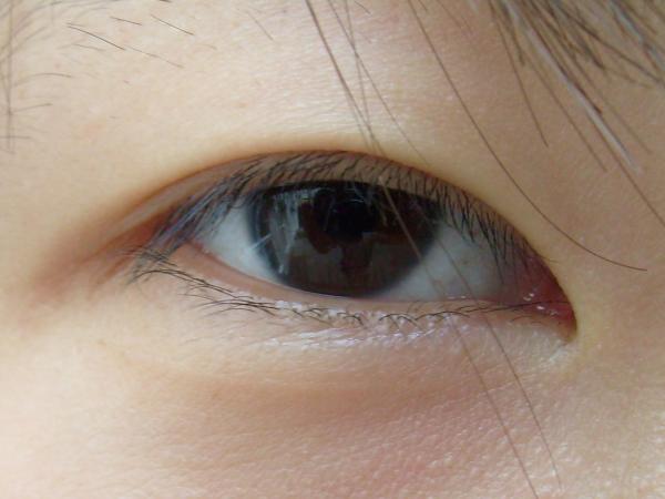
Blepharoplasty is eyelid surgery that removes or repositions excess skin, muscle, and fat of the upper or lower eyelids to restore function or improve appearance. Functional upper eyelid blepharoplasty is done when redundant skin (dermatochalasis) blocks the superior visual field or causes symptoms; cosmetic cases address ptosis of lash line, “bags,” or contour changes. Diagnosis is clinical, based on eyelid and brow measurements, photographs, and sometimes visual field testing. Treatment is surgical; outcomes depend on accurate diagnosis, technique selection, and ocular surface status.
Key Points
- Blepharoplasty is indicated functionally for visual field obstruction from upper lid dermatochalasis and cosmetically for contour concerns of the upper or lower lids.
- Preoperative evaluation must distinguish true ptosis and brow ptosis from excess skin; failure to address coexisting ptosis/brow descent leads to suboptimal results.
- Dry eye disease (ocular surface disease) increases risk of postoperative exposure symptoms; screening and optimization reduce complications.
- Serious complications are rare but include retrobulbar/orbital hemorrhage with vision loss; rapidly increasing pain, proptosis, or vision change after surgery requires immediate decompression.
- Lower eyelid malposition (retraction/ectropion) risk rises with preexisting laxity; prophylactic canthopexy/canthoplasty and conservative skin resection mitigate risk.
- Documentation (standardized photos and visual fields) is required for third-party coverage of functional upper lid blepharoplasty in the US.
Anatomy and Physiology
- Upper eyelid: skin, orbicularis oculi muscle, orbital septum, preaponeurotic fat (medial and central fat pads), levator aponeurosis and Müller muscle, tarsus. The eyelid crease forms where the levator aponeurosis inserts into the skin.
- Lower eyelid: thin skin, orbicularis, septum, fat compartments (medial, central, lateral), capsulopalpebral fascia, tarsus. Lateral canthal tendon stabilizes lid position.
- Age-related changes: thinning skin, lax septum with fat pseudoherniation, decreased levator tone or aponeurotic dehiscence, and canthal tendon laxity. These changes produce hooding, lash ptosis, and lower lid “bags.”
Etiology
- Involutional dermatochalasis with or without prolapse of orbital fat (most common).
- Eyelid laxity and canthal tendon weakening (involutional).
- Congenital eyelid crease variations (e.g., absent/low crease).
- Postinflammatory or edematous conditions leading to redundant skin (e.g., prior angioedema or blepharochalasis).
- Sequelae of chronic rubbing, contact dermatitis, or long-term steroid use.
- Thyroid eye disease with fat expansion (relative contraindication until stable).
Pathophysiology
- Redundant upper eyelid skin weighs on lashes and drapes over the pupil, reducing superior field and increasing frontalis muscle recruitment, causing brow ache and fatigue.
- Septal laxity allows anterior displacement of preaponeurotic (upper) or lower lid fat pads, creating contour bulges.
- Levator aponeurosis dehiscence (true ptosis) lowers the upper lid margin; blepharoplasty alone does not correct this and may unmask ptosis.
- Lower lid retraction/ectropion after aggressive skin/muscle resection or unaddressed laxity leads to exposure and tearing.
Epidemiology
- Blepharoplasty is among the most commonly performed facial surgeries in the US. Cosmetic “eyelid surgery” consistently ranks within the top 5 cosmetic surgical procedures annually, with over 100,000 cases reported in recent national statistics [1]. Functional blepharoplasty volume is substantial in Medicare-aged patients.
- Average age: mid-40s and older for involutional changes; younger adults for anatomic crease modification.
Classification
- By indication:
- Functional upper eyelid blepharoplasty (visual field obstruction, dermatochalasis-related symptoms).
- Cosmetic upper or lower eyelid blepharoplasty.
- By approach/technique:
- Upper eyelid: skin-only, skin–muscle, septal tightening, selective fat excision or repositioning, with or without ptosis repair or brow surgery.
- Lower eyelid: transconjunctival (fat excision/reposition; no skin incision) vs transcutaneous (subciliary/skin–muscle flap) with adjunctive canthopexy/canthoplasty; fat transposition to tear trough.
- By anatomic objective:
- Crease formation/augmentation (including “double eyelid” crease creation).
- Volume preservation vs reduction.
Symptoms and Signs
Functional upper eyelid dermatochalasis
- Symptoms: superior field loss (trouble seeing traffic lights or reading unless brow elevated), brow fatigue/ache, frontal headaches, dermatitis from skin-on-lash contact, lash ptosis.
- Signs: redundant skin overhanging lid margin, low or absent crease, makeup imprint on upper lid skin, reduced measured superior visual field that improves with eyelid taped, frontalis overaction, sometimes mild true ptosis.
Cosmetic upper eyelid concerns
- Symptoms: “heavy lids,” asymmetry, hollowing.
- Signs: dermatochalasis, variable preaponeurotic fat prolapse, low/absent crease, true ptosis or brow ptosis in some.
Cosmetic lower eyelid concerns
- Symptoms: under-eye “bags,” shadowing, festoons, tear-trough hollowing, perceived tired look.
- Signs: pseudoherniated fat pads, thin crepey skin, orbicularis hypertrophy, canthal laxity, midface descent, malar mounds/festoons.
Complications
- Early: ecchymosis/edema, chemosis, corneal abrasion, mild exposure symptoms, wound dehiscence, hematoma.
- Vision-threatening: retrobulbar/orbital hemorrhage with optic nerve compression; presents with severe pain, proptosis, decreased vision/afferent pupillary defect, elevated intraocular pressure; emergent lateral canthotomy/cantholysis is indicated [2,3].
- Intermediate/late:
- Overresection (lagophthalmos, hollow sulcus).
- Underresection/asymmetry.
- Lower lid retraction/ectropion, scleral show.
- New or worsened dry eye disease/exposure keratopathy.
- Diplopia from extraocular muscle injury (rare; inferior oblique during lower lid surgery).
- Infection, scarring, milia, pigmentary change.
- Levator dehiscence (iatrogenic ptosis) or unmasked preexisting ptosis.
- Very rare permanent vision loss after hemorrhage [3,4].
Diagnosis
Clinical evaluation
- History: visual field symptoms; headache or brow fatigue; contact lens wear; prior refractive surgery; dry eye disease; thyroid eye disease; anticoagulant/antiplatelet use; keloid history; systemic bleeding risk.
- Examination:
- External photos (frontal, oblique, upgaze/downgaze).
- Margin reflex distance 1 (MRD1), palpebral fissure height, levator function, brow position, eyelid crease height.
- Lash-to-fold distance and skin redundancy.
- Lower lid distraction/snapback test; lateral canthal position.
- Ocular surface: conjunctiva, cornea, tear film; Schirmer testing when indicated.
- Bell phenomenon, lagophthalmos risk.
- Counseling: distinguish dermatochalasis from true ptosis and brow ptosis; set realistic goals; discuss adjuncts (ptosis repair, brow lift, canthopexy).
Imaging and laboratory testing
- Not routinely required.
- Automated or Goldmann visual field testing with and without taping for functional upper eyelid cases to document impairment and improvement [5].
- Preoperative labs per anesthesia risk stratification; targeted thyroid testing if clinical suspicion of thyroid eye disease.
Differential diagnosis
| Entity | Distinguishing features |
|---|---|
| True blepharoptosis (levator dehiscence, myogenic, neurogenic) | Low lid margin (reduced MRD1) with normal/low skin; levator function reduced in myogenic; requires ptosis repair, not skin-only excision [6] |
| Brow ptosis | Low brow and forehead descent; improves superior field when brow elevated; may need brow lift |
| Blepharochalasis | Recurrent eyelid edema episodes leading to atrophic redundant skin; operate only after quiescence |
| Thyroid eye disease | Lid retraction, proptosis, restrictive strabismus; avoid cosmetic surgery until disease stable |
| Floppy eyelid syndrome | Lax lids, papillary conjunctivitis; treat OSA; higher risk of exposure postoperatively |
| Allergic or contact dermatitis | Pruritus, erythema, scaling; manage inflammation first |
| Chalazion | Focal tarsal nodule from meibomian gland; may mimic fullness; see chalazion page: https://myvisioncare.org/disease/chalazion |
| Preseptal/orbital cellulitis | Pain, erythema, fever; orbital signs in orbital cellulitis; urgent treatment |
| Conjunctivitis (bacterial) | Hyperemia, discharge, irritation; no structural eyelid excess; see bacterial conjunctivitis: https://myvisioncare.org/disease/bacterial-conjunctivitis |
| Keratitis | Pain, photophobia, corneal findings; not an eyelid contour issue; see keratitis: https://myvisioncare.org/disease/keratitis |
Treatment
Medical management
- Preoperative:
- Optimize ocular surface disease (lubricating drops/ointments; lid hygiene; consider punctal plugs or anti-inflammatory therapy as indicated) [7].
- Smoking cessation; coordinate anticoagulant/antiplatelet management with the prescribing clinician; avoid herbal supplements with bleeding risk [2].
- Postoperative:
- Cold compresses 10–15 minutes hourly while awake for 48 hours, then warm compresses as ecchymosis resolves.
- Lubrication: preservative-free artificial tears qid and ophthalmic ointment at bedtime for 1–2 weeks; more frequent if exposure symptoms occur.
- Topical ophthalmic antibiotic ointment (e.g., erythromycin 0.5% or bacitracin) to incisions bid–qid for 3–7 days; evidence for prophylaxis is limited; practice varies [2,6].
- Analgesia: acetaminophen; avoid NSAIDs immediately after surgery due to bleeding risk unless directed by surgeon.
- Activity: head elevation; avoid heavy lifting/strenuous activity 1–2 weeks; avoid eye makeup and contact lenses until incisions epithelialize (often 1–2 weeks).
Procedural and surgical management
-
Anesthesia: local with or without oral/IV sedation for most cases; general anesthesia for extensive revisions or combined procedures.
-
Upper eyelid blepharoplasty:
- Marking: crease placement individualized; skin pinch test to avoid overresection.
- Incision: within natural crease.
- Dissection: skin-only or skin–muscle flap; conservative excision of skin and/or orbicularis; open septum judiciously; selective medial/central fat excision or repositioning; levator/Müller inspection; concurrent ptosis repair if indicated.
- Closure: fine sutures; remove at 5–7 days.
-
Lower eyelid blepharoplasty:
- Transconjunctival approach: fat excision or transposition to tear trough; no external scar; preserves middle lamella; often combined with skin pinch for laxity.
- Transcutaneous (subciliary) approach: skin–muscle flap elevation; fat repositioning/excision; skin redraping; higher risk of retraction; often add lateral canthopexy/canthoplasty when laxity present.
- Volume-preserving strategies favored to avoid hollowing; address midface descent when relevant.
-
Adjuncts: brow lift, ptosis repair (levator advancement or Müller muscle-conjunctival resection), canthopexy/canthoplasty, resurfacing (peels/laser) for fine rhytids.
-
Coding (professional audience):
- ICD-10-CM: H02.83- Dermatochalasis of eyelid, with laterality/eyelid specificity.
- CPT: 15822/15823 (upper eyelid blepharoplasty; documentation of visual impairment for 15823), 15820/15821 (lower eyelid blepharoplasty).
Special populations
- Pediatric: Rare; generally limited to congenital crease anomalies or post-traumatic deformity; defer until growth allows stable planning unless functionally necessary.
- Pregnancy/lactation: Elective cosmetic surgery deferred; weigh anesthetic and medication exposure risks.
- Immunocompromised/diabetes: Higher infection and wound-healing risk; optimize glycemic control; meticulous asepsis.
- Elderly: Higher prevalence of dermatochalasis and laxity; evaluate cardiovascular/anticoagulation status; plan conservative resections.
- Thyroid eye disease: Wait for euthyroid state and disease quiescence; consider staged orbital decompression/strabismus procedures before lid surgery [6].
- Ocular surface disease/recent refractive surgery: Increased risk of exposure and dry eye exacerbation; aggressive lubrication and conservative tissue removal; preoperative counseling is essential [7].
Prognosis
- Symptom relief and aesthetic improvement rates are high when indications and technique are appropriate; recurrence reflects ongoing aging and individual tissue biology. Functional gains are durable; many patients maintain improved superior visual fields for years. Lower lid outcomes depend on addressing laxity and midface support. Severe complications are uncommon; permanent vision loss is rare [1–4,6]. Quantitative long-term durability data vary by technique.
Prevention and Patient Counseling
- Set goals: functional relief vs aesthetic refinement; discuss asymmetry tolerance and potential need for adjunct procedures.
- Review risks: bleeding, infection, scarring, asymmetry, dry eye worsening, lid malposition, vision changes; rare emergency of orbital hemorrhage.
- Postoperative care plan: cold compresses, elevation, lubrication, timed suture removal, activity restrictions, and sun protection for scars.
- Dry eye education: anticipate transient dryness/irritation; provide a dry eye care plan and refer to dry eye syndrome resources: https://myvisioncare.org/blog/dry-eye-syndrome
- Long-term: aging continues; revisions may be considered after adequate healing intervals if needed.
When to Seek Immediate Care
- Sudden vision loss, double vision, severe eye pain, or bulging of the eye.
- Rapidly increasing eyelid swelling or a firm, painful hematoma on one side.
- Inability to open the eye or new numbness of the eyelids/cheek.
- Persistent or bright-red bleeding that soaks dressings.
- Fever with spreading redness of the eyelids.
- Any corneal pain, light sensitivity, or discharge suggesting infection.
References
- American Society of Plastic Surgeons. 2022/2023 Plastic Surgery Statistics — Eyelid Surgery (Blepharoplasty) volumes. 2023. https://www.plasticsurgery.org/news/plastic-surgery-statistics.
- Codner MA, McCord CD. Upper and Lower Blepharoplasty. In: Neligan PC, ed. Plastic Surgery. 4th ed. Elsevier; 2017.
- AAO EyeWiki. Complications of Blepharoplasty. https://eyewiki.aao.org/Complications_of_Blepharoplasty
- Mahabir RC, et al. Orbital Hemorrhage After Blepharoplasty: Prevention and Treatment. Plast Reconstr Surg. 2011;127(2):716-722. doi:10.1097/PRS.0b013e3181fed6d3
- American Academy of Ophthalmology. Functional Indications for Upper Lid Blepharoplasty and Brow Ptosis Repair (Clinical Statement). 2017 update. https://www.aao.org/clinical-statement
- AAO EyeWiki. Upper Blepharoplasty; Lower Eyelid Blepharoplasty. https://eyewiki.aao.org/Upper_Blepharoplasty; https://eyewiki.aao.org/Lower_Eyelid_Blepharoplasty
- Stapleton F, et al. TFOS DEWS II: Impact of Iatrogenic Dry Eye. Ocul Surf. 2017;15(3):511-538. doi:10.1016/j.jtos.2017.05.003
This article is for informational purposes and is not a substitute for professional medical advice, diagnosis, or treatment. Always consult a qualified eye care professional about your specific condition. If you have sudden vision loss, severe eye pain, or an eye injury, seek emergency care immediately.