Dry eye syndrome is a chronic, multifactorial disorder of the tears and ocular surface that results in symptoms of discomfort, visual disturbance, and tear film instability with potential damage to the ocular surface. Most cases arise from meibomian gland dysfunction (evaporative dry eye) or reduced lacrimal tear production (aqueous-deficient dry eye). Cardinal symptoms include burning, grittiness, fluctuating blurry vision, and reflex tearing. Diagnosis is clinical, supported by tear breakup time, ocular surface staining, and targeted tests. Treatment is stepwise: tear supplements, eyelid hygiene and warm compresses, anti-inflammatory therapy (eg, cyclosporine or lifitegrast), meibomian gland therapies, punctal occlusion, and, in severe disease, serum tears or scleral lenses.
Key Points
- Dry eye syndrome results from tear film instability, hyperosmolarity, and ocular surface inflammation; neurosensory abnormalities may contribute.
- Meibomian gland dysfunction drives most evaporative dry eye; autoimmune lacrimal gland disease (eg, Sjögren syndrome) causes aqueous-deficient dry eye.
- Symptoms are often out of proportion to signs; fluctuating vision that clears after blinking suggests tear instability.
- Diagnosis is clinical; fluorescein tear breakup time < 10 seconds, corneal/conjunctival staining, abnormal Schirmer test, elevated tear osmolarity, and positive MMP-9 support the diagnosis.
- First-line therapy includes preservative-free artificial tears, environmental optimization, and eyelid hygiene; add anti-inflammatory agents and meibomian therapies as severity increases.
- Urgent evaluation is required for severe pain, marked light sensitivity, vision loss, a white corneal spot, or suspected infection.
Anatomy and Physiology
The tear film comprises:
- A superficial lipid layer from meibomian glands that retards evaporation and stabilizes the air–tear interface.
- An aqueous layer from the main and accessory lacrimal glands that provides moisture, nutrients, and antimicrobial proteins.
- A mucin layer from conjunctival goblet cells that promotes wetting of the corneal epithelium.
Blinking spreads the tear film and expresses meibum. Sensory input from the cornea and conjunctiva regulates basal and reflex tearing via trigeminal–parasympathetic pathways.
Etiology
-
Evaporative causes
- Meibomian gland dysfunction (MGD; obstructive or hyposecretory)
- Incomplete blinking, prolonged screen use
- Eyelid margin disease (eg, Demodex blepharitis, rosacea)
- Environmental: low humidity, air drafts, smoke, CPAP mask leak
- Topical medications and preservatives (eg, benzalkonium chloride)
-
Aqueous-deficient causes
- Autoimmune: primary or secondary Sjögren syndrome
- Lacrimal gland hypofunction (age-related, post-radiation, graft-versus-host disease)
- Neurotrophic deficiency, systemic dehydration
-
Mixed mechanisms are common.
-
Medication-related contributors
- Anticholinergics, antihistamines, antidepressants/SSRIs/SNRIs, beta-blockers, isotretinoin, diuretics.
-
Iatrogenic/exposure
- Contact lens wear, refractive surgery (eg, LASIK), eyelid malposition (lagophthalmos, ectropion).
Pathophysiology
A core mechanism is tear film instability leading to increased osmolarity, which triggers a cytokine-mediated inflammatory cascade, epithelial damage, and goblet cell loss. Meibomian gland obstruction alters lipid composition and increases evaporation. Aqueous deficiency reduces tear volume and clearance of inflammatory mediators. Chronic inflammation sustains a vicious cycle of instability and surface disease. Neurosensory abnormalities (hyperalgesia or hypoesthesia) can amplify symptoms or blunt protective reflexes.
Epidemiology
In US population-based data, approximately 6.8% of adults (about 16 million people) report a clinician diagnosis of dry eye disease; symptom prevalence is higher and increases with age and female sex [1]. Risk is elevated in autoimmune disease, rosacea, contact lens wearers, and post–refractive surgery patients [2].
Classification
-
By mechanism
- Aqueous-deficient dry eye
- Sjögren vs non-Sjögren
- Evaporative dry eye
- Intrinsic (MGD, blink disorders)
- Extrinsic (environmental, contact lenses, preservatives)
-
By severity (clinical staging; adapted from AAO/TFOS)
- Mild: episodic symptoms; minimal staining; TBUT mildly reduced.
- Moderate: frequent symptoms; conjunctival and corneal staining; reduced TBUT; early MGD signs.
- Severe: constant symptoms; filamentary keratitis, mucus strands; marked staining; conjunctival keratinization; keratitis complications.
Symptoms and Signs
-
Common symptoms
- Burning, stinging, foreign body sensation, dryness
- Fluctuating or blurry vision that improves after blinking
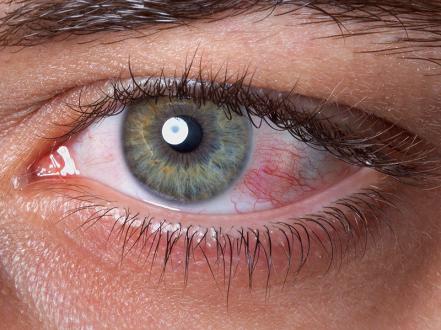
- Photophobia, ocular fatigue, redness
- Reflex tearing (epiphora) due to surface irritation
- Contact lens intolerance
-
Evaporative (MGD-predominant)
- Symptoms worsen with reading/screen use or wind exposure.
- Signs: thickened lid margins, telangiectasia, capped glands, turbid or inspissated meibum, foamy tear film, reduced TBUT.
-
Aqueous-deficient (eg, Sjögren)
- Symptoms often severe; associated dry mouth, dental caries, arthralgia, parotid enlargement.
- Signs: scant tear meniscus, reduced Schirmer, filamentary keratitis, diffuse conjunctival staining, mucus strands.
Complications
- Superficial punctate keratitis; filamentary keratitis
- Recurrent corneal erosions; persistent epithelial defects
- Secondary infectious keratitis and corneal ulceration (risk heightened with bandage lenses or immunosuppression) [2]
- Conjunctival scarring and keratinization in severe autoimmune disease
- Visual fluctuation affecting driving/reading; perioperative refractive surprises
Diagnosis
Clinical evaluation
-
History
- Symptom pattern, triggers (wind, screens), contact lens wear, systemic disease, medications.
- Symptom questionnaires (eg, Ocular Surface Disease Index [OSDI], Dry Eye Questionnaire-5 [DEQ-5]) help grade severity [2].
-
Examination
- External: blink rate/quality; lagophthalmos.
- Slit-lamp: tear meniscus height, debris, foamy tears; lid margin disease; meibomian gland expression quality.
- Tear film breakup time (TBUT) with fluorescein: < 10 seconds suggests instability.
- Corneal and conjunctival staining with fluorescein and lissamine green; document distribution and severity.
Imaging and laboratory testing
- Schirmer I (without anesthesia): ≤ 5 mm in 5 minutes supports aqueous deficiency.
- Tear osmolarity: ≥ 308 mOsm/L or intereye difference > 8 mOsm/L indicates instability/hyperosmolarity [2].
- Matrix metalloproteinase-9 (MMP-9) point-of-care test: positive suggests inflammation.
- Meibography: gland dropout or truncation supports MGD.
- Sjögren evaluation when suspected: anti-SSA/Ro, anti-SSB/La, ANA, RF; consider rheumatology referral and salivary gland biopsy per systemic guidelines [3].
Differential diagnosis
| Entity | Distinguishing features |
|---|---|
| Allergic conjunctivitis | Itching predominant; papillae; stringy mucus; seasonal exposure; responds to antihistamines/mast-cell stabilizers. |
| Blepharitis (anterior) | Lash crusting, collarettes; anterior lid margin inflammation; often coexists with MGD. |
| Bacterial conjunctivitis | Mucopurulent discharge, eyelids stuck in morning, papillary reaction; typically self-limited; see bacterial conjunctivitis. |
| Viral conjunctivitis (adenoviral) | Watery discharge, preauricular node, recent URI; highly contagious. |
| Exposure keratopathy | Lagophthalmos, nocturnal exposure, proptosis or facial nerve palsy; inferior staining. |
| Contact lens–related irritation | Symptoms tied to lens wear; punctate staining in lens zones; improve off lenses. |
| Medication toxicity (medicamentosa) | Chronic preserved drops; conjunctival hyperemia; punctate epitheliopathy. |
| Neuropathic ocular pain | Severe burning with minimal signs; hyperalgesia; comorbid migraine/fibromyalgia. |
| Corneal abrasion/ulcer | Acute pain, photophobia, focal infiltrate or epithelial defect; urgent evaluation; see keratitis. |
Treatment
Management is stepwise and mechanism-targeted, often combining approaches. Control surface inflammation and optimize meibomian function early; escalate if refractory.
Medical management
-
Education and environmental optimization
- Blink training; 20–20–20 breaks; humidifier; avoid direct airflow (fans, car vents); optimize workstation ergonomics.
- Adequate hydration; review and minimize contributory medications when feasible.
-
Tear supplementation
- Artificial tears (eg, carboxymethylcellulose, hyaluronate, polyethylene glycol/propylene glycol, HP-guar): 1–2 drops up to q2h as needed; prefer preservative-free if used > 4–6 times/day or in moderate–severe disease.
- Lipid-containing tears for MGD-predominant evaporative dry eye.
- Lubricating ointments at bedtime for nocturnal symptoms or exposure.
-
Eyelid hygiene and warm compresses
- Warm compresses 5–10 minutes once or twice daily; gentle lid massage to express meibum.
- Lid scrubs with dilute surfactant; targeted therapy for Demodex (tea tree oil–based lid wipes; lotilaner 0.25% ophthalmic solution is FDA-approved for Demodex blepharitis, not for dry eye per se).
-
Anti-inflammatory topical therapy
- Cyclosporine ophthalmic (eg, 0.05% one drop bid; 0.09% one drop q12h; 0.1% one drop bid): reduces T-cell–mediated inflammation; onset typically 1–3 months [2]. FDA-approved for dry eye disease.
- Lifitegrast 5% ophthalmic solution: one drop bid; LFA-1/ICAM-1 antagonist; symptomatic benefit may begin within 2–6 weeks; adverse effects include dysgeusia and irritation [2]. FDA-approved.
- Loteprednol etabonate 0.25% (Eysuvis): one drop qid for up to 2 weeks for acute signs and symptoms; monitor intraocular pressure with repeated courses [4]. FDA-approved.
- Short courses of other low-potency topical corticosteroids may be used off-label for flares; monitor for ocular hypertension and cataract.
-
Secretagogues and neuromodulation
- Varenicline solution nasal spray 0.03 mg/spray (Tyrvaya): 1 spray in each nostril bid to increase basal tear production via trigeminal parasympathetic pathway; nasal irritation and sneezing common [5]. FDA-approved.
- Oral pilocarpine 5 mg PO qid or cevimeline 30 mg PO tid may improve aqueous secretion in Sjögren; FDA-approved for xerostomia, used off-label for ocular dryness; cholinergic side effects and contraindications apply [2].
-
Meibomian gland–directed therapy
- Topical azithromycin 1% drops for lid margin disease (off-label for MGD); eyelid margin microblepharoexfoliation in-office.
- Oral tetracyclines for MGD/rosacea: doxycycline 20–50 mg PO bid (subantimicrobial) or 50–100 mg PO qd–bid for 4–12 weeks; consider maintenance at lower dose; photosensitivity and GI effects counsel [2].
-
Advanced/adjunctive therapies
- Autologous serum tears (eg, 20%–50% qid–8x/day) for severe ocular surface disease; off-label; compounding and infection control required [2].
- Scleral lenses/PROSE devices create a fluid reservoir improving optics and epithelial healing; infection prevention and care education essential [2].
- Moisture chamber goggles, nighttime eyelid taping or shields for exposure-related symptoms.
-
Nutritional supplements
- Omega-3 fatty acids showed no superiority to placebo in a large RCT (DREAM) for moderate–severe dry eye, so routine use for symptom relief is not supported by high-quality evidence [6].
Procedural and surgical management
- Punctal occlusion: temporary collagen or silicone plugs to reduce tear drainage; consider after controlling surface inflammation; thermal cautery for refractory cases [2].
- Thermal pulsation systems (eg, LipiFlow) and intense pulsed light (IPL) can improve meibomian gland function in selected patients; device-based therapies are adjuncts with variable availability and cost [2].
- Meibomian gland probing in obstructive MGD for symptom relief in experienced hands [2].
- Amniotic membrane (self-retained) for nonhealing epithelial defects or severe keratitis; tarsorrhaphy for exposure keratopathy.
- Bandage soft contact lenses may relieve pain in filamentary keratitis; concurrent antimicrobial prophylaxis and close monitoring are required due to infection risk.
Special populations
- Pediatric: Uncommon; consider congenital alacrima, ectodermal dysplasia, vernal/atopic disease, post–bone marrow transplant. Emphasize preservative-free lubricants, eyelid hygiene, and treat atopic comorbidity; subspecialty coordination often needed.
- Pregnancy/lactation: Symptoms may worsen; prioritize nonpharmacologic measures and preservative-free tears. Limited human data for cyclosporine and lifitegrast with minimal systemic absorption; use only if benefits outweigh risks; avoid systemic tetracyclines.
- Autoimmune/Sjögren and graft-versus-host disease: Early anti-inflammatory therapy, secretagogues, and escalation to serum tears or scleral lenses are often required; co-manage with rheumatology/hematology.
- Elderly: Polypharmacy review is essential; arthritis may limit drop instillation—consider delivery aids and ointments.
Prognosis
Dry eye syndrome is chronic with fluctuating severity. Most patients achieve symptomatic control with layered therapy and trigger avoidance, but relapse is common when treatment is reduced. Severe autoimmune or neurotrophic cases risk persistent epithelial disease and infectious keratitis; visual function improves when tear film stability is restored.
Prevention and Patient Counseling
- Reduce desiccating stressors: humidify indoor air, avoid direct airflow, wear wraparound eyewear outdoors.
- Schedule regular screen breaks and complete blinks.
- Perform daily eyelid hygiene and warm compresses for MGD.
- Use preservative-free tears when dosing frequently; avoid drops that “get the red out.”
- Discuss medication contributors with prescribing clinicians.
- Optimize contact lens wear schedules and hygiene; consider daily disposables or lens holidays during flares.
- Inform surgeons of dry eye before cataract or refractive surgery to optimize the ocular surface preoperatively.
When to Seek Immediate Care
- Sudden decrease in vision or a new white/gray spot on the cornea
- Severe eye pain or marked light sensitivity
- Thick discharge or eyelids stuck shut, especially with contact lens wear
- Chemical splash or foreign body in the eye
- Symptoms not improving after 2 weeks of appropriate care
References
- Dana R, Bradley JL, Guerin A, et al. Estimated Prevalence and Incidence of Dry Eye Disease in the United States. Am J Ophthalmol. 2019;202:47-54. doi:10.1016/j.ajo.2019.01.026.
- American Academy of Ophthalmology Cornea/External Disease Panel. Dry Eye Syndrome Preferred Practice Pattern. San Francisco, CA: AAO; 2023. https://www.aao.org/preferred-practice-pattern.
- Shiboski CH, Shiboski SC, Seror R, et al. 2016 ACR/EULAR Classification Criteria for Primary Sjögren’s Syndrome. Arthritis Rheumatol. 2017;69(1):35-45. doi:10.1002/art.39859.
- Eysuvis (loteprednol etabonate ophthalmic suspension) 0.25% [prescribing information]. Kala Pharmaceuticals, Inc.; 2020. https://www.accessdata.fda.gov/drugsatfda_docs/label/2020/211679s000lbl.pdf.
- Tyrvaya (varenicline solution) nasal spray [prescribing information]. Oyster Point Pharma, Inc.; 2021. https://www.accessdata.fda.gov/drugsatfda_docs/label/2021/213978s000lbl.pdf.
- Asbell PA, Maguire MG, Peskin E, et al; DREAM Study Research Group. n–3 Fatty Acid Supplementation for the Treatment of Dry Eye Disease. N Engl J Med. 2018;378(18):1681-1690. doi:10.1056/NEJMoa1709691.
- Stapleton F, Alves M, Bunya VY, et al. TFOS DEWS II Epidemiology Report. Ocul Surf. 2017;15(3):334–365. doi:10.1016/j.jtos.2017.05.003.
- Restasis (cyclosporine ophthalmic emulsion) 0.05% [prescribing information]. Allergan; 2017. https://www.accessdata.fda.gov/drugsatfda_docs/label/2017/050790s028lbl.pdf.
- Xiidra (lifitegrast ophthalmic solution) 5% [prescribing information]. Bausch + Lomb; 2023. https://www.accessdata.fda.gov/drugsatfda_docs/label/2023/208073s017lbl.pdf.
Disclaimer: This article is for informational purposes and is not a substitute for professional medical advice, diagnosis, or treatment. Always consult a qualified eye care professional about your specific condition. If you have sudden vision loss, severe eye pain, or an eye injury, seek emergency care immediately.