Hypermetropia (US: hyperopia), commonly called farsightedness, is a refractive error in which parallel light focuses behind the retina when accommodation is relaxed, causing blurred near vision and, in higher degrees, distance blur. It results most often from short axial length or insufficient corneal/lenticular power. Symptoms include eyestrain, frontal headaches, and near-vision difficulty; children may develop accommodative esotropia and amblyopia. Diagnosis is by refraction, preferably with cycloplegia in children. Treatment is optical correction with plus lenses; selected adults may be candidates for refractive surgery.
Key Points
- Hypermetropia (hyperopia) is a refractive error with image focus behind the retina; accommodation can mask distance blur until presbyopia.
- Children with moderate to high hypermetropia risk accommodative esotropia and amblyopia; cycloplegic refraction is essential.
- Optical correction with spectacles or contact lenses is safe and effective; full cycloplegic correction is standard in accommodative esotropia.
- Hyperopic laser vision correction (LASIK/PRK) is less predictable than myopic treatments and has higher regression; careful selection required.
- Hyperopes often have shallow anterior chambers and increased risk of primary angle-closure; gonioscopy is indicated when suspected.
- ICD-10-CM: H52.00–H52.03 (laterality-specific hypermetropia).
Anatomy and Physiology
Clear retinal focus requires coordinated optical power of the cornea and crystalline lens relative to the axial length of the eye. The cornea provides about two-thirds of refractive power; the crystalline lens fine-tunes focus and accommodates by increasing curvature via the ciliary muscle and zonules. In hypermetropia, the combined optical power is insufficient for eye length, placing the focal point posterior to the retina when accommodation is relaxed.
Etiology
- Axial: Short axial length (microphthalmos spectrum in extremes).
- Curvature: Flatter-than-average cornea or lens surfaces.
- Index: Reduced refractive index of lens (e.g., early cortical changes) or post-surgical states.
- Aphakia: Absence or dislocation of the crystalline lens.
- Functional: Accommodative insufficiency or paralysis unmasking latent hyperopia.
- Developmental/genetic: Familial aggregation; emmetropization delays or failures in infancy.
Pathophysiology
Accommodation can neutralize some hypermetropia (“facultative” component). Children possess robust accommodative amplitude, often maintaining clear distance vision at the cost of asthenopia and near blur. Persistent accommodative effort increases convergence via the accommodative convergence/accommodation (AC/A) link, predisposing to accommodative esotropia. With aging, presbyopia reduces accommodative reserve, revealing previously latent hyperopia and causing progressive near and distance blur. Shallow anterior chamber depth common in hyperopes elevates the risk for angle crowding and primary angle-closure.
Epidemiology
Hypermetropia is common at birth and decreases through early childhood with emmetropization. Clinically significant hyperopia persists in a subset of children and adults. Prevalence varies by age, race/ethnicity, and definition threshold; moderate-to-high hyperopia is a risk factor for accommodative esotropia and amblyopia [1–4]..
Classification
- By magnitude:
- Low (< +2.00 D), moderate (+2.00 to +5.00 D), high (> +5.00 D).
- By optical basis:
- Axial, curvature, index, aphakic.
- By accommodative status:
- Latent (masked by ciliary tone), manifest (detectable without cycloplegia), facultative (overcome by accommodation), absolute (not overcome by accommodation).
- By etiology:
- Simple (physiologic), pathological (associated with ocular anomalies), functional (accommodative paresis).
Symptoms and Signs
General hypermetropia
- Blurred near vision; distance blur with higher refractive error or fatigue.
- Asthenopia: eyestrain, frontal or brow ache, worse with prolonged near tasks.
- Intermittent diplopia or micropsia with accommodative spasm (rare).
- Reduced amplitude of accommodation relative to age norms.
- Reduced visual acuity without correction; improves with plus lenses.
Pediatric hypermetropia
- Avoidance of near tasks; inattention; head tilt.
- Intermittent or constant esodeviation at near (accommodative esotropia).
- Amblyopia risk: anisometropic or isoametropic.
- Normal external exam; esotropia may increase with accommodation.
Adults and presbyopic patients
- Progressive near blur in 40s–50s as presbyopia unmasks latent hyperopia.
- Need for higher plus adds for near; asthenopia with computer work.
- Angle-closure risk signs: narrow angles on gonioscopy; intermittent halos, brow ache.
Complications
- Accommodative esotropia.
- Amblyopia (anisometropic or isoametropic).
- Asthenopia impacting school/work performance.
- Primary angle-closure suspect (PACS), intermittent or acute primary angle-closure glaucoma in anatomically predisposed eyes.
- Aniseikonia and binocular vision symptoms if anisohyperopia is corrected asymmetrically.
Diagnosis
Clinical evaluation
- Visual acuity at distance and near (age-appropriate).
- Objective refraction:
- Cycloplegic retinoscopy/autorefraction in children and young adults; cyclopentolate 1% is commonly used (1–2 drops, 5 minutes apart; evaluate after 30–45 minutes) [2,5].
- Subjective refinement in cooperative patients after cycloplegia wears off or with fogging techniques.
- Binocular alignment and function: cover tests at distance/near, near point of convergence, AC/A estimation, stereopsis.
- Accommodation: amplitude (e.g., push-up), facility, and lag (dynamic retinoscopy) when symptoms suggest accommodative dysfunction.
- Anterior segment assessment: slit lamp; gonioscopy/AS-OCT if narrow angles suspected.
- Fundus examination to exclude structural causes (e.g., nanophthalmos, optic disc crowding).
Imaging and laboratory testing
- Keratometry/corneal topography for curvature assessment and refractive surgery planning.
- Ocular biometry (axial length, anterior chamber depth, lens thickness) for high hyperopia, surgical planning, and pathological microphthalmos.
- Anterior segment OCT or ultrasound biomicroscopy in angle-closure risk, plateau iris, or lens position assessment.
- No laboratory tests are indicated for uncomplicated hypermetropia.
Differential diagnosis
| Entity | Distinguishing features |
|---|---|
| Presbyopia | Age-related loss of accommodation; normal distance vision in emmetropes; plus add restores near only. |
| Myopia | Distance blur relieved by minus lenses; near often clear without correction. |
| Astigmatism | Ghosting/blur at all distances; improves with cylindrical correction. |
| Accommodative spasm | Variable refraction, miosis, near reflex overaction; fluctuating blur; cycloplegia reveals baseline. |
| Convergence insufficiency | Near diplopia, headaches; normal refraction; recedes with vergence therapy. |
| Dry eye disease | Fluctuating blur, burning, worse with screen use; signs on tear film testing; see dry eye syndrome. |
| Keratitis | Pain, photophobia, focal corneal opacity or staining; urgent evaluation for infection; see keratitis. |
| Bacterial conjunctivitis | Redness, purulent discharge, eyelids stuck; vision usually preserved; see bacterial conjunctivitis. |
| Chalazion | Localized lid nodule; induced astigmatism or localized blur if large; see chalazion. |
Treatment
Medical management
- Spectacles:
- Plus spherical lenses correct manifest hyperopia; in children, full cycloplegic correction is standard when esotropia or amblyopia risk is present [3,5].
- Options include single-vision distance with near add, bifocals, or progressive addition lenses in presbyopes or high AC/A esotropia.
- Contact lenses:
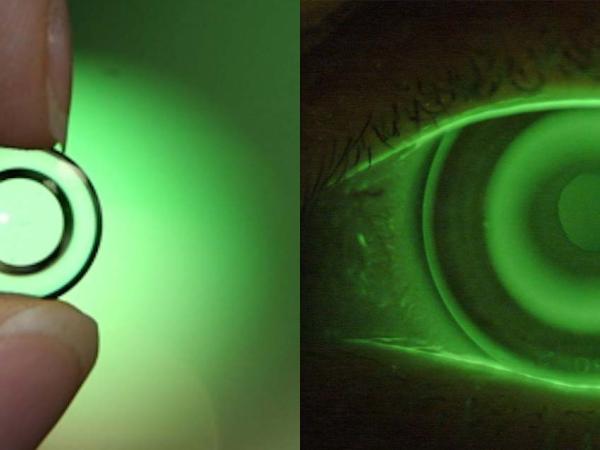
- Soft spherical or gas-permeable designs; multifocal options for presbyopic hyperopes.
- Counsel on hygiene; higher keratitis risk with overnight wear or poor lens care.
- Amblyopia therapy (when indicated):
- Occlusion (patching) or pharmacologic penalization (e.g., atropine 1% to the sound eye) per pediatric protocols, combined with refractive correction [3].
- Angle management:
- If anatomically narrow angles, consider prophylactic laser peripheral iridotomy per glaucoma guidelines; coordinate with glaucoma specialist.
Representative agents and use (clinician-directed):
- Cycloplegia for diagnostic refraction (off-label dosing details vary by age):
- Cyclopentolate 1%: 1–2 gtts, 5 minutes apart; assess after 30–45 minutes [2,5].
- Atropine 1% for home cycloplegia in select amblyopia/anomalous accommodation cases (specialist-supervised).
Procedural and surgical management
- Corneal laser vision correction (adults, stable refraction):
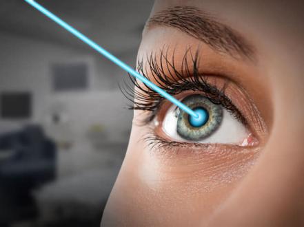
- Hyperopic LASIK and PRK can correct low-to-moderate hyperopia in suitable corneas; outcomes are less predictable than myopic treatments, with higher regression and risk of night-vision symptoms [6,7]. Typical treatable range is limited.
- Preoperative evaluation: stable refraction ≥12 months, adequate corneal thickness and diameter, normal topography/tomography, realistic expectations.
- Risks: dry eye exacerbation, glare/halos, under/overcorrection, ectasia (rare).
- Refractive lens exchange (RLE):
- Clear-lens extraction with IOL implantation can address high hyperopia or presbyopic needs; in patients without visually significant cataract, this is off-label refractive surgery [6].
- IOL selection: monofocal, bifocal/trifocal, or extended depth-of-focus; discuss dysphotopsias, need for readers, and potential anisometropia.
- Benefits include deepening of shallow anterior chamber in angle-closure suspects; risks include endophthalmitis, cystoid macular edema, retinal detachment (risk profile differs from myopes), and loss of accommodation.
- Historically used conductive keratoplasty:
- FDA-approved for low hyperopia/presbyopia in the past but largely abandoned due to regression and unpredictability [6,7].
Note: Phakic IOL options for hyperopia are limited in the US; currently available ICLs are approved for myopia and myopic astigmatism, not hyperopia [8].
Special populations
- Pediatric:
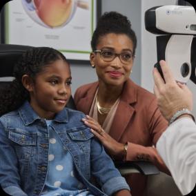
- Routine cycloplegic refraction for suspected hyperopia, strabismus, or amblyopia.
- Full correction for accommodative esotropia; prompt amblyopia therapy improves outcomes [3].
- Monitor emmetropization; prescribe earlier for high bilateral hyperopia or functional impairment.
- Pregnancy/lactation:
- Refractive surgery is deferred until several months postpartum; transient refractive and corneal changes can occur.
- Dry eye symptoms may worsen; prioritize spectacles/contacts with careful lens hygiene.
- Immunocompromised:
- Elevated infectious keratitis risk with contact lenses; prefer spectacles or stringent lens protocols; avoid overnight wear.
- Older adults:
- Presbyopia unmasks latent hyperopia; consider progressive lenses.
- Screen for narrow angles with gonioscopy where indicated; earlier lens extraction may be considered in angle-closure spectrum in coordination with glaucoma/cataract specialists.
Prognosis
Prognosis is excellent with appropriate optical correction. Children with hypermetropia corrected early have reduced risk of permanent amblyopia and strabismus. Hyperopic laser procedures can reduce dependence on glasses in selected adults, but outcomes may regress over time and enhancements may be needed [6,7]. Untreated significant hyperopia can cause chronic asthenopia, learning difficulties related to near tasks, accommodative esotropia, and amblyopia.
Prevention and Patient Counseling
- Refractive errors cannot be prevented; early detection and correction minimize complications.
- Counsel families about signs of accommodative esotropia and the need for adherence to spectacle wear and amblyopia therapy.
- Discuss realistic goals and limitations of refractive surgery for hypermetropia, including potential regression and visual side effects.
- Educate contact lens wearers on infection risks and proper care; avoid sleeping in lenses.
- Advise at-risk hyperopes about symptoms of angle-closure and the importance of periodic angle assessment.
When to Seek Immediate Care
- Sudden vision loss in one or both eyes.
- Severe eye pain, headache, halos around lights, nausea/vomiting (possible acute angle-closure).
- New constant eye crossing or double vision, especially in a child.
- Painful red eye with light sensitivity or discharge.
- After eye injury or chemical exposure.
References
- National Eye Institute. Facts About Refractive Errors. https://www.nei.nih.gov/learn-about-eye-health/eye-conditions-and-diseases/refractive-errors. Accessed July 24, 2026.
- American Academy of Ophthalmology. Pediatric Eye Evaluations Preferred Practice Pattern. Ophthalmology. 2022;129(1):P1–P151. doi:10.1016/j.ophtha.2021.09.017.
- American Association for Pediatric Ophthalmology and Strabismus (AAPOS). Accommodative Esotropia. https://aapos.org/glossary/accommodative-esotropia. Accessed July 24, 2026.
- EyeWiki. Hyperopia. American Academy of Ophthalmology. https://eyewiki.aao.org/Hyperopia. Accessed July 24, 2026.
- Donahue SP, Nixon CN; AAPOS Vision Screening Committee. Pediatric vision screening. Pediatrics. 2019;144(1):e20183912. doi:10.1542/peds.2018-3912.
- American Academy of Ophthalmology. Refractive Surgery Preferred Practice Pattern. Ophthalmology. 2022;129(11):P365–P476. doi:10.1016/j.ophtha.2022.07.012.
- Alio JL, Soria FA, Abbouda A, Panthier C. Laser in situ keratomileusis for hyperopia and hyperopic astigmatism: systematic review. J Cataract Refract Surg. 2019;45(2):167–182. doi:10.1016/j.jcrs.2018.10.036.
- U.S. Food and Drug Administration. LASIK Risks and Safety Information. https://www.fda.gov/medical-devices/lasik/what-are-risks. Accessed July 24, 2026.
ICD-10-CM
- H52.00 Hypermetropia, unspecified eye
- H52.01 Hypermetropia, right eye
- H52.02 Hypermetropia, left eye
- H52.03 Hypermetropia, bilateral
Disclaimer: This article is for informational purposes and is not a substitute for professional medical advice, diagnosis, or treatment. Always consult a qualified eye care professional about your specific condition. If you have sudden vision loss, severe eye pain, or an eye injury, seek emergency care immediately.