Keratoconus is a noninflammatory corneal ectatic disorder characterized by progressive stromal thinning and anterior protrusion, producing irregular astigmatism and decreased vision. Onset typically occurs in adolescence or early adulthood and is often bilateral but asymmetric. Diagnosis relies on corneal topography/tomography and pachymetry. Management ranges from spectacles and contact lenses to FDA‑approved corneal collagen cross-linking to slow or halt progression; advanced disease may require intracorneal ring segments or corneal transplantation.

Key Points

  • Keratoconus causes progressive corneal thinning and cone-shaped protrusion that induces irregular astigmatism and visual distortion.
  • Eye rubbing and atopic disease are strong modifiable risk factors; genetic predisposition is common.
  • Corneal topography/tomography is required to confirm early disease and monitor progression.
  • Corneal collagen cross-linking (epithelium-off) is FDA‑approved to slow progression; contact lenses improve vision but do not stop progression.
  • Acute corneal hydrops (sudden corneal edema from Descemet membrane rupture) is a sight-threatening complication requiring urgent care.
  • Pediatric keratoconus progresses faster; early detection and timely cross-linking improve long-term outcomes.

Anatomy and Physiology

The cornea is a 5-layer avascular structure (epithelium, Bowman layer, stroma, Descemet membrane, endothelium). The stroma (≈90% of thickness) comprises lamellae of type I collagen with interwoven architecture conferring tensile strength and shape stability. Keratocyte function, extracellular matrix cross-links, and intraocular pressure (IOP) together determine corneal biomechanics. Disruption of stromal collagen organization or cross-link density reduces biomechanical resistance, enabling ectasia.

Etiology

Keratoconus is multifactorial.

  • Genetic susceptibility: familial aggregation; complex polygenic inheritance; associations with connective tissue disorders (Ehlers–Danlos, Marfan) and Leber congenital amaurosis.
  • Environmental/behavioral: chronic eye rubbing (often related to atopy, vernal keratoconjunctivitis, allergic rhinitis, asthma), floppy eyelid syndrome, obstructive sleep apnea; contact lens overwear may exacerbate microtrauma.
  • Hormonal/physiologic modifiers: pregnancy may transiently worsen corneal biomechanics.

Pathophysiology

Biomechanical failure arises from decreased stromal collagen cross-linking, altered lamellar slippage, and keratocyte apoptosis. Oxidative stress and increased matrix metalloproteinase activity promote extracellular matrix degradation. Chronic mechanical trauma from eye rubbing amplifies local inflammatory mediators, further weakening the stroma. As ectasia progresses, anterior curvature steepens (especially inferiorly), the posterior surface bulges, and the cornea thins centrally or paracentrally. Acute corneal hydrops results from a focal Descemet membrane rupture with influx of aqueous into the stroma, causing sudden edema.

Epidemiology

  • Typical onset: puberty to age 30; progression often slows with age.
  • Bilateral in most patients but asymmetric in severity.
  • Prevalence estimates vary with diagnostic methods; population-based data suggest keratoconus is more common than historically reported with wider detection by Scheimpflug tomography [1], [2].
  • Higher prevalence reported in people with atopy and in certain ethnic populations (e.g., Middle Eastern, South Asian) [1].

Classification

  • By severity (Amsler–Krumeich): stage I–IV based on keratometry (K), spherical equivalent/myopia–astigmatism, corneal thickness, and scarring.
  • By phenotype:
    • Forme fruste keratoconus: subclinical changes without slit-lamp signs.
    • Keratoconus suspect: borderline topographic/tomographic indices without definitive ectasia.
    • Clinical keratoconus: slit-lamp signs and characteristic maps.
  • Complication subtype: acute corneal hydrops.

Symptoms and Signs

Typical/progressive keratoconus

  • Symptoms: progressive blurred and distorted vision, ghosting/diplopia, glare, halos, starbursts, increasing myopia and astigmatism, frequent prescription changes. Monocular symptoms predominate due to asymmetry.
  • Exam: irregular astigmatism on retinoscopy (scissoring), reduced best-corrected visual acuity, inferior/paracentral steepening on keratometry/topography, localized or generalized stromal thinning on pachymetry.
  • Slit-lamp signs: conical protrusion; Fleischer ring (iron deposition) at the cone base; Vogt striae (vertical stromal stress lines that disappear with gentle pressure); apical thinning and scarring; prominent corneal nerves; Munson sign (V-shaped lower lid with downgaze in advanced disease); Rizzuti phenomenon (conical light reflex on the nasal cornea).

Acute corneal hydrops

  • Sudden painful decrease in vision with tearing, photophobia, and marked corneal whitening from edema.
  • Large Descemet membrane break visible on slit-lamp with anterior segment OCT; epithelial bullae may be present.

Complications

  • Apical scarring reducing visual potential.
  • Acute corneal hydrops; risk of corneal perforation is low but vision may decline substantially.
  • Contact lens intolerance; contact lens–associated keratitis if hygiene is poor.
  • Iatrogenic ectasia after refractive surgery in undetected keratoconus or suspects.
  • Postoperative issues: high or irregular astigmatism after keratoplasty; graft rejection (penetrating keratoplasty more than deep anterior lamellar keratoplasty).

Diagnosis

Clinical evaluation

  • History: onset age, progression, atopy, eye rubbing, family history, contact lens wear, pregnancy status, sleep position and OSA symptoms.
  • Visual acuity, manifest/cycloplegic refraction.
  • Slit-lamp biomicroscopy for classic signs and scarring; evaluate for vernal/atopic disease.
  • Retinoscopy for irregular reflex.
  • Intraocular pressure (may read falsely low on Goldmann applanation due to thin cornea).

Imaging and laboratory testing

  • Placido-disc topography: anterior surface curvature; inferior–superior asymmetry; asymmetric bow-tie with skewed axes.
  • Scheimpflug tomography (e.g., Pentacam) or dual Scheimpflug–Placido systems: anterior/posterior elevation maps, pachymetry distribution, Belin/Ambrosio Enhanced Ectasia Display for early detection.
  • Anterior segment OCT: epithelial thickness mapping (epithelial compensation patterns), stromal and Descemet visualization; confirms hydrops and Descemet breaks.
  • Ultrasound pachymetry if needed.
  • Corneal biomechanics (e.g., Corvis ST, Ocular Response Analyzer) adjunctive in suspects, not diagnostic alone.
  • Laboratory testing is not routine; consider for suspected associated connective tissue disorders based on systemic signs.

Differential diagnosis

Entity Distinguishing features
Pellucid marginal degeneration Inferior peripheral thinning (4–8 o’clock) with “crab-claw” pattern; ectasia just above thinned band; normal central thickness.
Keratoglobus Generalized corneal thinning and globular protrusion; onset often earlier; high risk of perforation with trauma.
Post-LASIK/PRK ectasia History of keratorefractive surgery; inferior/posterior steepening; thin residual stromal bed.
Contact lens warpage Topographic irregularity resolves after lens holiday; epithelial mapping supports warpage.
Terrien marginal degeneration Superior peripheral thinning with lipid deposition and vascularization; against-the-rule astigmatism; little inflammation.
Irregular astigmatism from scars Localized topographic distortion correlates with scar; stable over time.
Infectious keratitis Acute pain, focal infiltrate with overlying epithelial defect, anterior chamber reaction; positive staining and cultures.

Treatment

Medical management

  • Optical correction:
    • Spectacles or soft toric contact lenses in early disease with regular astigmatism.
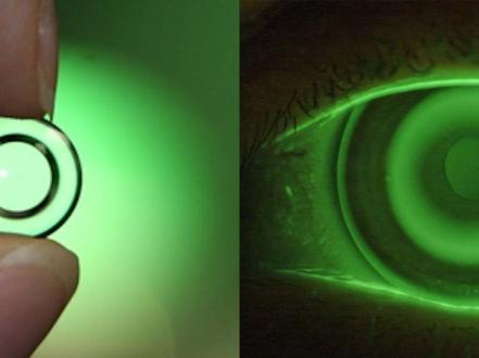
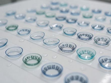
    • Rigid gas-permeable (RGP), hybrid, piggyback (soft under RGP), or scleral/prosthetic replacement of the ocular surface ecosystem (PROSE) lenses for irregular astigmatism. These improve optics but do not halt progression.
  • Atopy and eye rubbing control:
    • Topical dual-action antihistamine/mast cell stabilizers (e.g., olopatadine 0.1% bid or 0.2% qd; ketotifen 0.025% bid) to reduce itch; short, supervised courses of topical corticosteroids (e.g., loteprednol 0.2% qid, taper) for vernal flares; cold compresses; lubricants.
    • Counsel to avoid eye rubbing; consider nighttime eye shields or taping if sleep-related rubbing.
  • Dry eye optimization to improve contact lens tolerance (preservative-free tears; punctal occlusion when indicated).
  • Acute corneal hydrops:
    • Hypertonic sodium chloride 5% drops qid and ointment at bedtime to reduce edema; cycloplegic (e.g., cyclopentolate 1% tid) for pain/photophobia.
    • Topical corticosteroid (e.g., prednisolone acetate 1% qid) to mitigate inflammation; prophylactic topical antibiotic if epithelial defects (e.g., moxifloxacin 0.5% qid).
    • Bandage contact lens for epithelial bullae.
    • Intracameral gas tamponade (e.g., SF6 or C3F8) with/without compression sutures can hasten resolution and reduce scarring in large Descemet breaks [3].

FDA/regulatory note: No topical drug reverses keratoconus. Cross-linking is a procedural therapy approved as below.

Procedural and surgical management

  • Corneal collagen cross-linking (CXL), epithelium-off (epi-off):
    • FDA‑approved Photrexa (riboflavin 5′‑phosphate) solutions with KXL UVA device for progressive keratoconus and post‑refractive ectasia [4].
    • Standard (Dresden) parameters: riboflavin saturation after epithelial removal, then UVA 365 nm at 3 mW/cm² for 30 minutes (total 5.4 J/cm²). Variants (accelerated) exist; long-term equivalence data are evolving [3].
    • Minimum stromal thickness target commonly ≥400 μm after de-epithelialization; hypotonic riboflavin may be used to swell thin corneas [1].
    • Epi-on and transepithelial protocols are investigational/off-label in the US.
  • Intracorneal ring segments (ICRS, e.g., Intacs):
    • FDA‑approved for keratoconus to flatten the cone and reduce irregular astigmatism in selected cases with clear central cornea and adequate thickness. May improve spectacle or contact lens tolerance; does not stop progression [5].
  • Topography‑guided surface ablation combined with CXL:
    • Off‑label; in highly selected cases, limited photoablation to regularize the corneal surface followed by CXL may improve optics. Candidacy requires adequate corneal thickness and careful risk assessment.
  • Keratoplasty:
    • Deep anterior lamellar keratoplasty (DALK) preferred when endothelium is healthy; preserves endothelium, reduces rejection risk, and yields good tectonic and optical outcomes.
    • Penetrating keratoplasty (PK) for full‑thickness scarring, severe hydrops scarring, or when DALK is not feasible. Lifelong risk of graft rejection; high/irregular postoperative astigmatism is common and may still require rigid or scleral lenses.
  • Postoperative and longitudinal care:
    • Lifelong monitoring with topography/tomography after CXL or surgery.
    • Refit contact lenses as shape stabilizes; manage ocular surface.

Coding notes: ICD‑10‑CM keratoconus includes H18.6x series. CPT/Category III for CXL and CPT for DALK/PK vary by year and payer policy.

Special populations

  • Pediatric: Faster progression, higher risk of scarring; low threshold for early tomography and for proceeding to CXL once progression is documented. General anesthesia may be required for young or developmentally delayed patients.
  • Pregnancy/lactation: Keratoconus may worsen during pregnancy; defer elective refractive or corneal reshaping procedures. Safety of CXL in pregnancy/lactation is not established; generally postponed until postpartum unless vision-threatening progression.
  • Down syndrome and neurodiverse patients: High prevalence and frequent eye rubbing; behavioral strategies, protective eyewear during sleep, and caregiver education are central. Consider anesthesia planning for procedures.
  • Elderly: Natural stiffening may slow progression; monitor for contact lens intolerance and scarring–related visual limits.

Prognosis

Without intervention, many patients experience progressive irregular astigmatism and visual decline over years. With timely CXL, most eyes stabilize structurally, preserving or improving functional vision with optical correction [3]. Advanced disease may still require ICRS or keratoplasty. Long-term follow-up with serial tomography is essential to detect late progression or postoperative changes.

Prevention and Patient Counseling

  • Avoid eye rubbing; treat allergic eye disease aggressively to control itch.
  • Adhere to contact lens hygiene and replacement schedules; avoid overnight wear unless lenses are prescribed for that purpose.
  • Schedule regular eye examinations with corneal imaging, especially during adolescence and early adulthood, or if atopy/eye rubbing persists.
  • Discuss family screening when keratoconus is diagnosed, given familial risk.
  • Post-CXL: protect the eye until re-epithelialization; avoid water exposure and contact lens wear until cleared.

When to Seek Immediate Care

  • Sudden drop in vision, severe eye pain, or corneal whitening/swelling.
  • Large corneal blister, new light sensitivity, or tearing that does not improve.
  • Red, painful eye with discharge while wearing contact lenses.
  • After cross-linking or surgery: increasing pain, persistent blurred vision, or discharge.
  • Any eye injury to a known thin or protruding cornea.

References

  1. EyeWiki. Keratoconus. American Academy of Ophthalmology. Updated 2024. https://eyewiki.aao.org/Keratoconus
  2. National Eye Institute. Keratoconus. Updated 2022. https://www.nei.nih.gov/learn-about-eye-health/eye-conditions-and-diseases/keratoconus
  3. Cochrane Eyes and Vision Group. Corneal collagen cross-linking for keratoconus. Cochrane Database Syst Rev. 2021;[Issue unknown]:[pages]. doi:
  4. U.S. Food and Drug Administration. Summary of Safety and Effectiveness: Photrexa Viscous, Photrexa and KXL System (PMA P150034). 2016. https://www.fda.gov
  5. U.S. Food and Drug Administration. INTACS Prescription Inserts for Keratoconus (P980040/Sxxx). https://www.fda.gov
  6. StatPearls. Keratoconus. Treasure Island (FL): StatPearls Publishing; 2023. https://www.ncbi.nlm.nih.gov/books/NBK470415/
  7. AAO Preferred Practice Pattern. Corneal Ectasia (Keratoconus and Related Ectasias). American Academy of Ophthalmology; 2022. https://www.aao.org/preferred-practice-pattern
  8. Gomes JA, Tan D, Rapuano CJ, et al. Global consensus on keratoconus and ectatic diseases. Cornea. 2015;34(4):359‑369. doi:10.1097/ICO.0000000000000408
  9. Reinhart WJ, Musch DC, Jacobs DS, et al. Deep anterior lamellar keratoplasty as an alternative to penetrating keratoplasty: a report by the AAO. Ophthalmology. 2011;118(1):209‑218. doi:10.1016/j.ophtha.2010.11.002

ICD‑10‑CM: H18.60 Keratoconus, unspecified; H18.6x subcodes by stability/laterality.

Disclaimer: This article is for informational purposes and is not a substitute for professional medical advice, diagnosis, or treatment. Always consult a qualified eye care professional about your specific condition. If you have sudden vision loss, severe eye pain, or an eye injury, seek emergency care immediately.