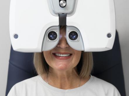
Strabismus is a misalignment of the eyes in which the visual axes do not point at the same target. It may be constant or intermittent and may manifest as inward, outward, vertical, or torsional deviation. Children typically present with eye turning and reduced depth perception; adults often present with double vision (diplopia). Diagnosis is clinical using cover testing and prism measurements plus cycloplegic refraction. Management includes refractive correction, amblyopia therapy, prisms, chemodenervation, and extraocular muscle surgery.
Key Points
- Strabismus is an ocular misalignment that can be comitant (angle constant in all gaze positions) or incomitant (angle varies, often neurologic or restrictive).
- Early detection and treatment in children reduces risk of amblyopia (functional vision loss) and improves stereopsis.
- Cycloplegic refraction is essential; accommodative esotropia commonly corrects with full hyperopic glasses, sometimes with a near add.
- Adults with new-onset diplopia require prompt evaluation to exclude cranial nerve palsy, thyroid eye disease, or stroke.
- Treatment is stepwise: optical correction and amblyopia therapy first; prisms or botulinum toxin in selected cases; surgery for persistent deviations.
- Success often requires long-term follow-up; some patients need more than one procedure.
Anatomy and Physiology
Six extraocular muscles per eye coordinate binocular alignment and pursuit/saccadic movements: medial, lateral, superior, and inferior recti; superior and inferior obliques. Innervation is via cranial nerves III (oculomotor), IV (trochlear), and VI (abducens). Sensory fusion in the visual cortex integrates near-simultaneous, matched retinal images into single binocular vision and stereopsis. Disruption of motor alignment, sensory fusion, or both produces misalignment, suppression, or diplopia.
Etiology
- Refractive/accommodative: uncorrected hyperopia (accommodative esotropia), high AC/A (accommodative convergence to accommodation) ratio.
- Congenital/infantile forms: large-angle comitant esotropia presenting in early infancy.
- Sensory: unilateral reduced vision from cataract, corneal scar, retinal disease.
- Neurologic: cranial nerve III, IV, VI palsies; brainstem/cerebellar lesions; skew deviation.
- Restrictive: thyroid eye disease (thyroid-associated ophthalmopathy), orbital fracture with muscle entrapment, scarring.
- Decompensated phoria: breakdown of tonic/binocular control with fatigue, illness, or age.
- Systemic associations: prematurity, periventricular leukomalacia, cerebral palsy, Down syndrome, fetal alcohol spectrum disorders.
- Iatrogenic: postoperative after retinal surgery, botulinum toxin spread, local anesthesia myotoxicity.
Pathophysiology
Misalignment arises from imbalance of muscle forces or neural signals, impaired fusional vergence, or abnormal sensory input. In children, persistent misalignment leads to cortical suppression of the deviating eye and development of amblyopia and loss of stereopsis. In adults with established binocular vision, acquired misalignment typically produces diplopia. Long-standing deviations can cause muscle length-tension adaptation (contracture) and secondary deviations.
Epidemiology
An estimated 2% to 4% of U.S. children have manifest ocular misalignment, with esotropia and exotropia most common [1,2]. Amblyopia affects approximately 1% to 3% of children and often coexists with misalignment [1]. Adult-onset deviations occur from decompensated phorias, microvascular cranial neuropathies (diabetes, hypertension), thyroid eye disease, trauma, and neurodegenerative disease [3].
Classification
- By direction: esotropia (inward), exotropia (outward), hypertropia/hypotropia (vertical), cyclotropia (torsional).
- By constancy: constant vs intermittent.
- By gaze dependency: comitant vs incomitant.
- By onset: infantile (early), childhood, adult-acquired.
- By mechanism:
- Accommodative (fully or partially) vs nonaccommodative.
- Sensory vs motor.
- Paralytic (neurogenic) vs restrictive.
- Specific entities: infantile esotropia, accommodative esotropia (with/without high AC/A), intermittent exotropia (basic, divergence excess, convergence insufficiency), vertical deviations (superior oblique palsy), convergence insufficiency.
Symptoms and Signs
Infantile/early esotropia
- Large, constant inward deviation presenting in first 6 months of life.
- Cross fixation, latent nystagmus, dissociated vertical deviation; minimal diplopia due to suppression.
- Limited abduction may mimic sixth nerve palsy but full abduction on doll’s head maneuver supports comitancy.
Accommodative esotropia (with/without high AC/A)
- Onset in toddlers/preschoolers; worse at near if high AC/A ratio.
- Improved or straight with full hyperopic correction; amblyopia risk in one eye.
Intermittent exotropia
- Outward drift, especially with distance fixation, fatigue, inattention, or bright light (closing one eye outdoors).
- Variable control; reduced stereoacuity at distance.
Vertical deviations (e.g., superior oblique palsy)
- Head tilt away from involved side, vertical/torsional diplopia in adults.
- Positive Bielschowsky head tilt test; hypertropia increases in contralateral gaze and ipsilateral head tilt.
Sensory deviations
- Large-angle, often constant deviation in the eye with reduced acuity; poor fusion potential.
Neurogenic or restrictive deviations
- Incomitant, gaze-limited; diplopia in involved directions.
- Ptosis, anisocoria (III palsy), abduction deficit (VI palsy), inelasticity and pain in thyroid eye disease, or motility restriction after trauma.
Common findings across types:
- Asymmetric corneal light reflex (Hirschberg), cover-uncover and alternate cover tests show tropia magnitude and control.
- Reduced stereopsis; amblyopia signs in children (fixation preference, poor vision without organic cause).
Complications
- Amblyopia (functional monocular vision loss) in children.
- Loss of stereopsis and fine depth perception.
- Suppression scotoma development; anomalous retinal correspondence.
- Diplopia (particularly in adults) causing reading difficulty, driving hazards, and falls.
- Abnormal head posture leading to neck/back strain.
- Psychosocial effects: reduced self-esteem, social stigma.
- Contracture of rectus muscles and secondary deviations over time.
Diagnosis
Clinical evaluation
- History: age at onset, variability, diplopia, trauma, neurologic symptoms, systemic disease, family history, prior treatment.
- Visual acuity (age-appropriate), fixation behavior, and monocular occlusion responses.
- Alignment:
- Corneal light reflex (Hirschberg/Krimsky).
- Cover-uncover (detect manifest deviation) and prism and alternate cover test (quantify angle) at distance/near, with and without correction.
- Ocular motility in nine gaze positions; duction/version limits; head tilt testing.
- Sensory testing: stereopsis (Randot, Titmus), Worth 4-dot; suppression/diplopia assessment.
- Cycloplegic refraction (cyclopentolate 1% for children >6 months; 0.5% for infants) to identify hyperopia, anisometropia, astigmatism.
- Anterior/posterior segment examination to exclude sensory causes (media opacity, retinal disease).
- Neurologic screening: pupils, ptosis, cranial nerves, cerebellar signs.
Imaging and laboratory testing
- Indications: acute cranial neuropathy signs, noncomitant deviations without clear cause, orbital trauma, restrictive signs, neurologic symptoms.
- Modalities:
- MRI brain/orbits (cranial nerve palsy, demyelination, mass).
- CT orbits (fracture, entrapment, calcification) and active thyroid eye disease.
- Laboratory tests guided by suspicion (e.g., thyroid function for thyroid-associated ophthalmopathy; glucose/HbA1c and blood pressure for microvascular neuropathies).
Differential diagnosis
| Entity | Distinguishing features |
|---|---|
| Pseudoesotropia (epicanthal folds, flat nasal bridge) | Normal cover tests; symmetric corneal light reflex; appearance improves with age |
| Decompensated phoria | Intermittent symptoms, asthenopia; small angle on alternate cover; normal motility |
| Convergence insufficiency | Exodeviation greater at near; receded near point of convergence; symptomatic while reading |
| Cranial nerve VI palsy | Limited abduction; incomitant esotropia worse at distance and toward affected side; new-onset diplopia in adults |
| Cranial nerve IV palsy | Hypertropia with torsion; positive head tilt test; vertical diplopia |
| Thyroid eye disease | Restrictive motility with pain/tightness; proptosis, lid retraction; variable diplopia |
| Orbital floor fracture (entrapment) | Pain, limitation of upgaze; positive forced ductions; infraorbital hypesthesia |
| Keratitis/uveitis with photophobia | Red, painful eye; decreased vision; no comitant deviation; corneal or anterior segment inflammation present |
| Bacterial conjunctivitis | Conjunctival injection and discharge; no true misalignment on cover testing |
Treatment
Medical management
- Optical correction
- Full cycloplegic hyperopic correction for accommodative esotropia; consider bifocal add (+2.00 to +3.00 D) for high AC/A ratio to align at near [4].
- Address anisometropia and astigmatism to optimize acuity and fusion potential.
- Fresnel or ground-in prisms for small, comitant deviations and symptomatic diplopia, particularly in adults.
- Amblyopia therapy (children)
- Occlusion (patching) of the dominant eye: typical regimens include 2 hours/day for moderate amblyopia and up to 6 hours/day for severe, with near activities to stimulate the amblyopic eye [5,6].
- Pharmacologic penalization: atropine 1% to the sound eye (daily or weekend dosing) is comparable to patching for moderate amblyopia in children 3–7 years [5]. Avoid in narrow angles and monitor for systemic anticholinergic effects.
- Chemodenervation
- Botulinum toxin type A injection to selected extraocular muscles can be used for acute cranial nerve palsies, infantile esotropia adjunctive therapy, and small-angle deviations in adults; dosing and technique are specialist-determined [7]. Some uses are off-label depending on indication; the product labeling includes treatment of strabismus in adults.
- Vision therapy/orthoptics
- Office- and home-based convergence exercises are beneficial for primary convergence insufficiency to reduce symptoms and improve near point of convergence [8].
- Underlying condition management
- Optimize thyroid eye disease, diabetes, hypertension; treat sensory causes (e.g., cataract).
Note: Systemic medications do not correct ocular misalignment; therapy targets optical, sensory, chemodenervation, or surgical pathways.
Procedural and surgical management
- Indications: persistent cosmetically or functionally significant deviation despite optimal optical/amblyopia therapy; symptomatic diplopia; abnormal head posture; documented sensory benefit potential.
- Procedures:
- Horizontal muscle surgery (recession/resection/plication) for esotropia/exotropia; bilateral or unilateral approaches based on angle and pattern. Adjustable sutures are advantageous in adults for intra/postoperative refinement.
- Vertical muscle surgery for hypertropia, including inferior oblique weakening for overaction, superior oblique tuck for lax tendon, or rectus vertical recessions.
- Transposition procedures for paralytic strabismus (e.g., vertical rectus transposition for sixth nerve palsy).
- Scar release and recessions for restrictive disease after quiescence (thyroid eye disease) and/or orbital decompression when indicated.
- Chemodenervation as primary or adjunct in selected cases.
- Perioperative considerations:
- General anesthesia in children; local anesthesia with sedation often feasible in adults.
- Risks: undercorrection/overcorrection, need for reoperation, diplopia (transient or persistent), slipped muscle, anterior segment ischemia (risk rises with multiple rectus muscles), infection, scarring.
- Coding (examples; verify payer specifics):
- CPT 67311 (strabismus surgery, 1 horizontal muscle), 67312 (2 muscles), 67314 (1 vertical muscle), 67335 (adjustable suture), 67320 (transposition).
Special populations
- Pediatric
- Timely amblyopia therapy is critical; most plasticity exists before age 7–8 years, but treatment can benefit older children.
- Earlier alignment (often within the first 6–24 months) for infantile esotropia improves likelihood of some stereopsis [2,4].
- Screen siblings; strong familial aggregation exists.
- Pregnancy/lactation
- Prefer nonpharmacologic amblyopia strategies; defer elective chemodenervation or surgery until postpartum when feasible. Atropine and other anticholinergics cross placenta and into breast milk; use only if benefits outweigh risks.
- Immunocompromised/anticoagulated
- Standard care with attention to infection prophylaxis and perioperative anticoagulation management per systemic guidelines.
- Older adults
- Address fall risk and driving safety; prisms or occlusion foils can palliate diplopia if surgery is deferred.
Prognosis
Outcomes depend on cause, age at onset, and timeliness of treatment. Fully accommodative esotropia typically aligns with proper spectacles; partially accommodative cases may need surgery. Intermittent exotropia has variable natural history; some remain stable, others progress. Adult paralytic deviations due to microvascular neuropathies often improve over 3–6 months; persistent misalignment may require prisms, chemodenervation, or surgery. Multiple operations may be necessary over a lifetime to maintain alignment and binocular function [2,3].
Prevention and Patient Counseling
- Promote pediatric vision screening at least once in all children ages 3–5 years to detect amblyopia or its risk factors, per USPSTF [9].
- Correct significant refractive errors early.
- Adhere to patching/atropine regimens; missed therapy reduces visual potential.
- Control systemic risk factors (diabetes, thyroid disease).
- Adults with new diplopia should avoid driving until evaluated and wear an occlusion patch or opaque foil temporarily if diplopia is disabling.
When to Seek Immediate Care
- Sudden double vision, especially with headache, droopy eyelid, unequal pupils, or weakness or numbness in the face or limbs.
- New eye misalignment after head or eye injury.
- Painful, red eye with light sensitivity and blurred vision.
- Rapidly worsening bulging eyes, severe eye movement pain, or decreased vision in thyroid disease.
- Any sudden vision loss.
References
- National Eye Institute. Facts About Strabismus. NIH/NEI. Accessed 2026. https://www.nei.nih.gov/learn-about-eye-health/eye-conditions-and-diseases/strabismus
- American Academy of Ophthalmology. Esotropia and Exotropia Preferred Practice Pattern. San Francisco, CA: AAO; 2022 update. https://www.aao.org/preferred-practice-pattern
- American Academy of Ophthalmology. Adult Strabismus Preferred Practice Pattern. San Francisco, CA: AAO; 2020 update. https://www.aao.org/preferred-practice-pattern
- AAPOS. Strabismus in Children (Patient Information). American Association for Pediatric Ophthalmology and Strabismus. https://aapos.org/glossary/strabismus
- Pediatric Eye Disease Investigator Group. A randomized trial of atropine vs patching for treatment of moderate amblyopia in children. N Engl J Med. 2002;347(9): 621-629. doi:10.1056/NEJMoa021397.
- Pediatric Eye Disease Investigator Group. A randomized trial of prescribed patching regimens for moderate amblyopia in children. Arch Ophthalmol. 2003;121(5): 603-611. doi:10.1001/archopht.121.5.603.
- BOTOX (onabotulinumtoxinA) injection, for intramuscular use. Full Prescribing Information. FDA/Allergan. Revised 2023. https://dailymed.nlm.nih.gov/dailymed/drugInfo.cfm?setid=cd674305-4e48-4c54-8b6d-61542a76e08f
- Scheiman M, et al. Randomized clinical trial of treatments for symptomatic convergence insufficiency in children. Arch Ophthalmol. 2005;123(1):14-24. doi:10.1001/archopht.123.1.14.
- US Preventive Services Task Force. Vision Screening in Children Ages 3 to 5 Years: US Preventive Services Task Force Recommendation Statement. JAMA. 2017;318(9):836-844. doi:10.1001/jama.2017.11260. [Check for 2023 reaffirmation/update]
- StatPearls. Strabismus. Treasure Island (FL): StatPearls Publishing; updated 2024. https://www.ncbi.nlm.nih.gov/books/NBK554592/
This article is for informational purposes and is not a substitute for professional medical advice, diagnosis, or treatment. Always consult a qualified eye care professional about your specific condition. If you have sudden vision loss, severe eye pain, or an eye injury, seek emergency care immediately.