Viral conjunctivitis is inflammation of the conjunctiva—the thin mucous membrane covering the white of the eye and the inside of the eyelids—caused most commonly by adenoviruses. It presents with acute red eye, watery discharge, foreign body sensation, and often a follicular (bumpy) tarsal conjunctival reaction with preauricular lymphadenopathy. Diagnosis is clinical; laboratory testing is rarely needed. Management is primarily supportive; topical corticosteroids are reserved for severe cases with membranes or visually significant corneal involvement.
Key Points
- Adenoviruses cause the majority of viral conjunctivitis in adults and are highly contagious for 10–14 days from symptom onset [1][2].
- Hallmarks include watery discharge, conjunctival follicles, and tender preauricular lymph nodes; epidemic keratoconjunctivitis (EKC) can cause subepithelial corneal infiltrates and photophobia.
- Treatment is supportive: cold compresses, artificial tears, and topical antihistamine/mast-cell stabilizers for itch; topical corticosteroids are used selectively for membranes or visually significant keratitis (off-label).
- Topical antibiotics and anti-adenoviral antivirals are not indicated; avoid corticosteroids if herpes simplex virus (HSV) is suspected.
- Strict hygiene and temporary exclusion from work/school during active tearing/crusting reduce transmission; contact lens wear should be stopped until fully resolved.
- Consider alternate diagnoses when pain, photophobia, or decreased vision are prominent (e.g., keratitis, uveitis, acute glaucoma).
Anatomy and Physiology
The conjunctiva comprises a non-keratinized stratified epithelium and a substantia propria rich in lymphoid tissue, blood vessels, and goblet cells. Viral pathogens trigger a follicular response—aggregates of lymphocytes in the tarsal conjunctiva. Corneal epithelial involvement in adenoviral infection can progress to anterior stromal immune-mediated subepithelial infiltrates that affect quality of vision.
Etiology
- Adenoviruses (most common): nonspecific follicular conjunctivitis, pharyngoconjunctival fever (PCF), and epidemic keratoconjunctivitis (EKC) [1][2].
- Enterovirus 70 and coxsackievirus A24 variant: acute hemorrhagic conjunctivitis (AHC) [3].
- Herpes simplex virus (HSV-1): unilateral follicular conjunctivitis, blepharoconjunctivitis, dendritic keratitis.
- Varicella-zoster virus (VZV): conjunctivitis with dermatomal vesicular rash (herpes zoster ophthalmicus).
- Molluscum contagiosum: chronic follicular conjunctivitis due to eyelid lesions shedding virus into the tear film.
- Less common: measles, rubella, mumps, SARS-CoV-2 (usually mild conjunctival hyperemia) [4].
Pathophysiology
Adenoviruses infect conjunctival and corneal epithelial cells, inciting a robust innate and adaptive immune response. In EKC, epithelial keratitis may appear within days, followed by immune-mediated subepithelial infiltrates 1–2 weeks after onset that can persist for months. Pseudomembranes form from fibrin and inflammatory exudate on the tarsal conjunctiva and can cause scarring if not removed. Viral shedding from conjunctival secretions facilitates rapid spread via direct contact and fomites.
Epidemiology
- Viral conjunctivitis is the most common cause of infectious conjunctivitis in adults in the United States; adenovirus accounts for most cases [1][2].
- Outbreaks occur in community settings (schools, offices) and healthcare facilities; PCF has been associated with swimming pools [1][2].
- Incubation typically 5–12 days; patients remain contagious while tearing/crusting persists, typically 10–14 days from onset [2].
Classification
- Adenoviral conjunctivitis
- Nonspecific follicular conjunctivitis
- Pharyngoconjunctival fever (PCF): conjunctivitis, fever, sore throat, preauricular nodes
- Epidemic keratoconjunctivitis (EKC): severe conjunctivitis with corneal involvement (epithelial keratitis, subepithelial infiltrates), membranes/pseudomembranes
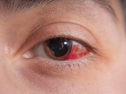
- Acute hemorrhagic conjunctivitis (AHC): enterovirus/coxsackievirus; subconjunctival hemorrhage, pain, photophobia
- Herpetic conjunctivitis (HSV, VZV): often unilateral; may include vesicular lid lesions, keratitis
- Molluscum-related chronic follicular conjunctivitis
Symptoms and Signs
-
General viral conjunctivitis
- Acute onset redness, watery/serous discharge, burning/foreign body sensation
- Follicular tarsal reaction, chemosis (conjunctival edema)
- Tender preauricular lymphadenopathy
- Usually starts unilateral and becomes bilateral within 1–3 days
-
Pharyngoconjunctival fever (PCF)
- Fever, pharyngitis, headache, malaise
- Follicular conjunctivitis with watery discharge and tender preauricular nodes
-
Epidemic keratoconjunctivitis (EKC)
- Marked chemosis, eyelid edema, photophobia
- Pseudomembranes on tarsal conjunctiva; bleeding on peeling
- Corneal epithelial punctate keratitis followed by subepithelial infiltrates (SEIs) 1–2 weeks in, causing photophobia, glare, and reduced contrast sensitivity
-
Acute hemorrhagic conjunctivitis (AHC)
- Sudden onset pain, photophobia, lid edema
- Scattered petechiae and subconjunctival hemorrhages
- Highly contagious; rapid community spread
-
Herpetic conjunctivitis (HSV/VZV)
- Often unilateral
- Vesicular lid lesions (blepharitis), dendritic epithelial keratitis (HSV) or pseudodendrites (VZV)
- Decreased corneal sensation possible
-
Molluscum-related
- Chronic unilateral/bilateral follicular conjunctivitis
- Umbilicated eyelid skin lesion(s) with lash line involvement
Complications
- Subepithelial corneal infiltrates with photophobia and visual disturbance (EKC)
- Pseudomembrane scarring, symblepharon (adhesions)
- Persistent dry eye symptoms after acute infection
- Secondary bacterial infection (uncommon)
- Misdiagnosis of HSV leading to inappropriate corticosteroid use with worsening keratitis
Diagnosis
Clinical evaluation
- Diagnosis is clinical in most cases based on history (recent sick contacts, exposure) and examination showing watery discharge, follicles, and preauricular adenopathy.
- Fluorescein staining may reveal punctate epithelial erosions in adenoviral keratitis or dendrites in HSV.
- Red flags suggesting alternate or additional pathology: moderate to severe pain, marked photophobia, decreased vision, corneal opacity, hypopyon, copious purulent discharge, contact lens wear, or trauma.
Imaging and laboratory testing
- Not routinely required.
- Point-of-care adenoviral antigen tests exist but have variable sensitivity; negative results do not exclude adenoviral disease.
- PCR testing is reserved for atypical cases, severe outbreaks, immunocompromised hosts, or infection control investigations [1].
- Obtain microbiologic testing if hyperacute purulent conjunctivitis suggests gonococcus or if chlamydial conjunctivitis is suspected.
Differential diagnosis
| Entity | Distinguishing features |
|---|---|
| Allergic conjunctivitis | Intense itching, bilateral, stringy/mucoid discharge, papillary reaction, history of atopy; no preauricular nodes |
| Bacterial conjunctivitis | Mucopurulent discharge, lids stuck on awakening, rapid response to topical antibiotics; follicles uncommon; see our page on bacterial conjunctivitis |
| Keratitis (infectious, contact lens-related) | Pain, photophobia, corneal infiltrate/ulcer, decreased vision; urgent evaluation; see our page on keratitis |
| Anterior uveitis (iritis) | Photophobia, consensual pain, ciliary flush, cells/flare in anterior chamber |
| Acute angle-closure glaucoma | Severe pain, halos, mid-dilated fixed pupil, corneal edema, markedly elevated intraocular pressure |
| HSV blepharoconjunctivitis | Unilateral, vesicular lid lesions, dendritic epithelial lesions with terminal bulbs on fluorescein staining, reduced corneal sensation |
| VZV conjunctivitis | Dermatomal vesicular rash (V1), pseudodendrites without terminal bulbs, often older adults or immunocompromised |
| Molluscum contagiosum | Chronic follicular conjunctivitis with eyelid umbilicated papules along lid margin/lashes |
| Dry eye disease | Fluctuating vision, burning, worse with screens/airflow, minimal discharge; chronic course |
| Nasolacrimal duct obstruction (pediatric) | Tearing and discharge since infancy, worse with colds; pressure over sac may express discharge |
Treatment
Medical management
-
Supportive care (first-line for adenoviral conjunctivitis)
- Preservative-free artificial tears qid to q2h for comfort.
- Cold compresses several times daily.
- Topical antihistamine/mast-cell stabilizers for itch and chemosis: ketotifen 0.025% 1 drop bid (OTC), olopatadine 0.1% 1 drop bid or 0.2% 1 drop daily.
- Oral analgesics (e.g., acetaminophen, NSAIDs) as needed.
-
Topical corticosteroids (off-label; selective use)
- Indications: pseudomembranes/membranes, severe inflammation with functional impairment, or visually significant corneal SEIs.
- Options: loteprednol etabonate 0.5% 1 drop qid, or fluorometholone 0.1% 1 drop qid, then taper over 1–2 weeks as symptoms improve.
- Avoid or defer if HSV keratitis is suspected or not excluded. Use the lowest effective dose and shortest duration to limit prolonged viral shedding [1][5].
-
Pseudomembrane management
- Careful mechanical peeling at the slit lamp after topical anesthesia to reduce scarring and symblepharon formation, followed by a short course of topical corticosteroid.
-
Antivirals/antibiotics
- No approved topical antiviral is effective against adenoviral conjunctivitis; cidofovir is not used clinically due to toxicity [1].
- Routine topical antibiotics are not indicated; reserve for clear bacterial superinfection.
-
Povidone-iodine (PVI) drench (off-label)
- In-office 5% PVI irrigation/scrub early in disease may reduce viral load and shorten symptom duration in some studies; evidence base is limited and heterogeneous [6]. Consider in EKC outbreaks or severe early presentations after risk–benefit discussion.
-
Herpetic conjunctivitis (HSV/VZV)
- Manage per herpetic eye disease protocols; if epithelial keratitis is present, topical ganciclovir 0.15% gel 5 times daily until healing, then tid for 7 days, or oral acyclovir/valacyclovir regimens [5]. Avoid corticosteroids without antiviral coverage.
-
Contact lens wear
- Discontinue lenses during active disease; discard or disinfect lenses and cases before resuming wear after full resolution.
Procedural and surgical management
- Membrane/pseudomembrane removal at the slit lamp with cotton-tipped applicator or forceps after topical anesthesia; consider hemostasis as needed.
- Debridement of molluscum lesions causing chronic conjunctivitis (curettage or cryotherapy) resolves the conjunctival inflammation.
- No role for surgery in routine adenoviral conjunctivitis.
Special populations
-
Pediatric
- PCF is more common. Exclude from daycare/school while eyes are actively tearing or matted; children can generally return when discharge and matting have resolved and they can maintain hygiene. Antibiotics are not required for return since disease is viral [2].
- Neonates with conjunctivitis require urgent evaluation to exclude gonococcal, chlamydial, or herpetic disease.
-
Pregnancy/lactation
- Supportive care preferred. If corticosteroids are necessary for severe keratitis/membranes, use the lowest effective potency and duration. Systemic antivirals for herpetic disease require obstetric coordination.
-
Immunocompromised
- May have prolonged shedding and more severe keratoconjunctivitis; consider confirmatory testing (PCR) and closer follow-up. Manage herpetic disease with systemic antivirals and ID co-management as indicated.
-
Older adults
- Coexisting dry eye may worsen symptoms; favor preservative-free tears and environmental modifications.
Prognosis
Uncomplicated adenoviral conjunctivitis resolves within 1–3 weeks. Corneal SEIs in EKC can persist for months and cause photophobia and fluctuating blur but usually improve with time; some cases require intermittent topical steroids or off-label topical calcineurin inhibitors. AHC resolves within 3–7 days. Recurrence is uncommon but reinfection with a different adenoviral serotype can occur.
Prevention and Patient Counseling
- Hand hygiene with soap and water or alcohol-based rubs; avoid touching/rubbing eyes.
- Do not share towels, pillows, cosmetics, or eye drops.
- Disinfect commonly touched surfaces (phones, keyboards).
- Avoid swimming pools and close-contact sports while actively symptomatic.
- Stay home from work/school while eyes are red with tearing/crusting; resume when discharge and matting have resolved and hygiene can be maintained [2].
- In clinics: strict instrument disinfection effective against nonenveloped viruses (e.g., diluted bleach), single-use tonometer tips when possible, and cohorting/isolation during outbreaks [1].
Link to related content:
- See differences from bacterial conjunctivitis on our page: bacterial conjunctivitis.
- Corneal involvement raises concern for keratitis; see our keratitis page.
- Residual irritation after infection may overlap with dry eye; see our overview of dry eye syndrome.
- Recurrent eyelid bumps are not viral conjunctivitis; see our chalazion page.
When to Seek Immediate Care
- Severe eye pain, sensitivity to light, or sudden decrease in vision
- Symptoms in a contact lens wearer (risk of corneal infection)
- Marked eyelid swelling with copious pus
- A newborn or infant with a red, draining eye
- A painful red eye with a new facial rash or blisters (possible shingles)
- History of eye trauma or chemical exposure
- Worsening symptoms after a few days of home care
References
- AAO EyeWiki. Adenoviral Conjunctivitis. American Academy of Ophthalmology. Updated 2023. https://eyewiki.aao.org/Adenoviral_Conjunctivitis
- Centers for Disease Control and Prevention (CDC). Adenoviruses: Clinical Overview. Accessed 2026. https://www.cdc.gov/adenovirus/hcp/clinical-overview.html
- Chang CH, et al. Acute hemorrhagic conjunctivitis. N Engl J Med. 2020;382(22):2167. doi:10.1056/NEJMicm1911841
- Sen M, et al. Ocular manifestations of COVID-19. Surv Ophthalmol. 2021;66(5):669-675. doi:10.1016/j.survophthal.2021.02.001
- American Academy of Ophthalmology. Conjunctivitis Preferred Practice Pattern. 2023. https://www.aao.org/preferred-practice-pattern/conjunctivitis-ppp
- Trinavarat A, Atchaneeyasakul L. Povidone-iodine in adenoviral conjunctivitis: a randomized clinical trial. J Med Assoc Thai. 2012;95 Suppl 4:S124-30.
- Azari AA, Barney NP. Conjunctivitis: a systematic review. JAMA. 2013;310(16):1721-1729. doi:10.1001/jama.2013.280318
- Tabbara KF. Ocular complications of adenoviral infections. Curr Opin Ophthalmol. 2001;12(6):467-471. doi:10.1097/00055735-200112000-00011
- StatPearls. Adenoviral Conjunctivitis. Updated 2023. https://www.ncbi.nlm.nih.gov/books/NBK470271/
Reviewed: July 2026 Last updated: July 2026
By: [ASSIGN] Medical Reviewer: [ASSIGN]
This article is for informational purposes and is not a substitute for professional medical advice, diagnosis, or treatment. Always consult a qualified eye care professional about your specific condition. If you have sudden vision loss, severe eye pain, or an eye injury, seek emergency care immediately.