Anterior uveitis is inflammation of the anterior uveal tract—primarily the iris and ciliary body—causing ocular pain, photophobia, redness, and decreased vision. It is most often idiopathic or associated with systemic autoimmune disease, and less commonly due to infection, trauma, or drug exposure. Diagnosis is clinical by slit-lamp biomicroscopy showing anterior chamber cells and flare; ancillary testing is guided by history and exam. Treatment typically includes topical corticosteroids and cycloplegics, with targeted therapy for infectious or systemic causes and close monitoring for complications.

Key Points

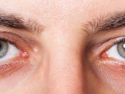
  • Anterior uveitis (iritis/iridocyclitis) presents with eye pain, photophobia, ciliary flush, and a small, sluggish pupil; slit-lamp shows cells and flare.
  • Most cases are noninfectious and idiopathic or HLA-B27–associated; rule out syphilis and consider sarcoidosis, tuberculosis, and herpes viruses when suspected.
  • Initial therapy is topical corticosteroids plus cycloplegics; taper steroids slowly to prevent rebound inflammation.
  • Complications include posterior synechiae, cataract, glaucoma, and cystoid macular edema; intraocular pressure must be monitored.
  • Avoid prostaglandin analogs and miotics during active inflammation; treat infectious uveitis with pathogen-directed therapy alongside cautious anti-inflammatory treatment.
  • Recurrent disease is common, especially in HLA-B27–associated cases; coordinate care for underlying systemic disease.

Anatomy and Physiology

The uvea comprises the iris, ciliary body, and choroid. In anterior uveitis, inflammation primarily involves the iris and anterior ciliary body. Breakdown of the blood–aqueous barrier permits leukocytes and protein (cells and flare) to enter the anterior chamber. Inflammatory debris may deposit on the corneal endothelium as keratic precipitates; posterior synechiae form when the inflamed iris adheres to the lens.

Etiology

  • Noninfectious, immune-mediated
    • HLA-B27 spondyloarthropathies: ankylosing spondylitis, reactive arthritis, psoriatic arthritis, inflammatory bowel disease
    • Juvenile idiopathic arthritis (JIA; often chronic, insidious, and asymptomatic)
    • Sarcoidosis
    • Behçet disease
  • Infectious
    • Herpes simplex virus (HSV), varicella-zoster virus (VZV)
    • Cytomegalovirus (CMV; typically hypertensive, recurrent anterior uveitis in immunocompetent adults)
    • Syphilis (Treponema pallidum)
    • Tuberculosis (Mycobacterium tuberculosis)
    • Less common: Lyme disease, toxoplasmosis (rarely isolated anterior), others based on geography/exposures
  • Trauma and surgery
    • Blunt/penetrating trauma, postoperative inflammation
    • Lens-induced uveitis (phacoantigenic/phacolytic)
  • Drug-induced
    • Cidofovir, rifabutin, bisphosphonates, checkpoint inhibitors; topical prostaglandin analogs may rarely precipitate uveitis
  • Distinct clinical entities
    • Fuchs heterochromic iridocyclitis (low-grade chronic)
    • Posner–Schlossman (glaucomatocyclitic crisis; recurrent with high IOP)
  • Masquerade syndromes
    • Intraocular lymphoma, retained lens fragments

Pathophysiology

Antigen-driven inflammation disrupts tight junctions in iris vasculature and nonpigmented ciliary epithelium, allowing inflammatory cells and proteins into aqueous humor. Cytokine-mediated trabeculitis impairs aqueous outflow, elevating intraocular pressure (IOP). Fibrin and posterior synechiae can occlude the pupil (seclusio pupillae), causing iris bombe and secondary angle closure. Chronic inflammation promotes cataract formation, band keratopathy (calcium deposition in Bowman layer), and cystoid macular edema (CME) via perifoveal capillary leakage.

Epidemiology

Anterior uveitis is the most common anatomic subtype of uveitis in the United States, comprising the majority of uveitis cases seen in ophthalmic practice [1], [2]. HLA-B27–associated acute anterior uveitis accounts for a substantial subset of recurrent unilateral cases in adults [2]. JIA-associated chronic anterior uveitis is a leading cause of visual morbidity in children [3]. Precise incidence and prevalence estimates vary by region and methodology.

Classification

Standardization of Uveitis Nomenclature (SUN) categories apply [4]:

  • By location: anterior (iris/ciliary body)
  • By onset: sudden vs insidious
  • By duration: limited (<3 months) vs persistent (≥3 months)
  • By course: acute (single episode), recurrent (≥2 episodes separated by ≥3 months of inactivity), chronic (relapses <3 months after stopping treatment)
  • By phenotype: granulomatous (large “mutton-fat” keratic precipitates, iris nodules) vs nongranulomatous (fine KPs)
  • By laterality: unilateral, alternating unilateral, or bilateral
  • By etiology: infectious vs noninfectious vs masquerade

Symptoms and Signs

Acute nongranulomatous anterior uveitis (often HLA-B27–associated)

  • Symptoms: ocular pain, photophobia (including consensual), redness, tearing, blurred vision.
  • Signs: ciliary flush, anterior chamber cells and flare, fine keratic precipitates, small sluggish pupil, posterior synechiae, variable IOP (often low early; elevated if trabeculitis).

Granulomatous anterior uveitis (e.g., sarcoidosis, TB)

  • Symptoms: subacute onset, mild pain/photophobia.
  • Signs: large “mutton-fat” KPs, iris nodules (Koeppe at pupillary margin; Busacca in stroma), vitritis may be present.

Herpetic anterior uveitis (HSV/VZV)

  • Symptoms: unilateral recurrent episodes, pain, photophobia.
  • Signs: sectoral iris atrophy, elevated IOP (trabeculitis), decreased corneal sensation, keratitis (dendrites or pseudodendrites in VZV) may coexist.

CMV anterior uveitis

  • Symptoms: recurrent unilateral blurred vision, halos.
  • Signs: marked IOP spikes, fine-to-medium KPs (often coin-shaped), minimal pain; responds poorly to steroids alone.

Fuchs heterochromic iridocyclitis

  • Symptoms: often asymptomatic until cataract; minimal photophobia.
  • Signs: diffuse stellate KPs, heterochromia, absence of posterior synechiae, low-grade cells, vitreous opacities, secondary glaucoma.

Posner–Schlossman syndrome

  • Symptoms: recurrent episodes of halos and blurred vision with mild discomfort.
  • Signs: very high IOP with mild anterior chamber inflammation; open angles between attacks.

JIA-associated chronic anterior uveitis

  • Symptoms: often none; detected on screening.
  • Signs: chronic cells/flare, band keratopathy, posterior synechiae, cataract, amblyopia risk.

Complications

  • Posterior synechiae; seclusio pupillae; iris bombe with secondary angle-closure glaucoma
  • Ocular hypertension and secondary open-angle glaucoma (trabeculitis, steroid response)
  • Cataract (inflammatory and steroid-induced)
  • Cystoid macular edema (leading cause of vision loss in uveitis)
  • Band keratopathy
  • Hypotony (ciliary body shutdown) in severe chronic disease
  • Corneal edema (from elevated IOP or endothelial dysfunction)

Diagnosis

Clinical evaluation

  • History: onset/course, laterality, recurrence; systemic symptoms (back pain, psoriasis, bowel disease, oral/genital ulcers, rash, cough/dyspnea, fevers), infectious risks (STIs, TB exposure), prior herpes zoster ophthalmicus, medications (rifabutin, bisphosphonates), trauma or surgery, pediatric rheumatologic history.
  • Examination:
    • Visual acuity; external and pupillary exam (miosis, posterior synechiae)
    • Slit-lamp: anterior chamber cell/flare (grade per SUN), KPs (type/distribution), iris nodules, fibrin, hypopyon
    • IOP measurement (avoid applanation over active epithelial defects)
    • Dilated fundus exam to assess for vitritis, CME, posterior segment involvement

Imaging and laboratory testing

Testing is selective, directed by phenotype and history; avoid indiscriminate “shotgun” panels.

  • Baseline or high-yield tests when indicated:
    • Syphilis serology: treponemal (e.g., FTA-ABS or TPPA) plus nontreponemal (RPR or VDRL) [5]
    • HLA-B27 in recurrent acute nongranulomatous anterior uveitis without another diagnosis [2]
    • Chest imaging (X-ray; CT if needed) and serum ACE/lysozyme when sarcoidosis suspected [2]
    • TB testing (IGRA such as QuantiFERON-TB Gold; chest imaging) if risk factors or granulomatous uveitis [2]
    • ANA in children at risk for JIA-associated uveitis [3]
    • HSV/VZV/CMV aqueous PCR in atypical, hypertensive, or steroid-refractory cases [2]
  • Ancillary ocular imaging:
    • Optical coherence tomography (OCT) for CME
    • Fluorescein angiography when CME or vasculitis suspected

Differential diagnosis

Entity Distinguishing features
Bacterial conjunctivitis Diffuse conjunctival injection, mucopurulent discharge, minimal photophobia, normal pupil/IOP, no anterior chamber cells; often both eyes
Viral conjunctivitis Watery discharge, preauricular node, follicles, no anterior chamber cells
Keratitis (infectious) Corneal epithelial defect/ulcer, focal stromal infiltrate, significant photophobia; positive corneal staining
Episcleritis Sectoral redness, mild discomfort, blanches with phenylephrine; no photophobia
Scleritis Severe deep, boring pain; scleral tenderness; violet hue; may not blanch; often systemic autoimmune disease
Acute angle-closure glaucoma Severe pain, halos, headache/nausea, mid-dilated fixed pupil, corneal edema, very high IOP; shallow anterior chamber
Endophthalmitis Postoperative/postinjection context or trauma; severe pain, hypopyon, vitritis, profound vision loss
Corneal abrasion History of trauma/contact lens; epithelial defect; pain improves with topical anesthetic in clinic (not for home use)

Related conditions for patient context: chalazion (/disease/chalazion), bacterial conjunctivitis (/disease/bacterial-conjunctivitis), and keratitis (/disease/keratitis) can also cause a red eye but have different exam findings.

Treatment

Goals are to control inflammation, relieve pain/photophobia, prevent synechiae, treat underlying causes, and monitor for complications.

Medical management

  • Topical corticosteroids (first-line for noninfectious anterior uveitis)
    • Prednisolone acetate 1%: 1 drop q1–2h while awake for 24–48 hours, then taper based on response (e.g., qid → bid → qd over weeks)
    • Difluprednate 0.05%: 1 drop qid (four times daily) for moderate–severe disease; taper carefully; higher risk of IOP rise [DailyMed/FDA label, 6]
    • Loteprednol 0.5–1%: for milder cases or steroid responders
    • Monitor IOP at baseline and during treatment
  • Cycloplegic/mydriatic agents (pain relief; prevent synechiae)
    • Cyclopentolate 1%: 1 drop tid (three times daily)
    • Homatropine 2–5%: 1 drop bid–tid (2–3 times daily)
    • Atropine 1%: 1 drop qd–bid (daily to twice daily) for severe inflammation or fibrin
  • IOP management
    • Aqueous suppressants: timolol 0.5% bid; brimonidine 0.1–0.2% tid; dorzolamide 2% tid; consider oral acetazolamide 250–500 mg PO qid short term for marked spikes (avoid in pregnancy and sulfonamide allergy)
    • Avoid prostaglandin analogs and miotics during active uveitis (may exacerbate inflammation)
  • Antiviral therapy (for confirmed/suspected herpetic etiologies; use with topical steroids)
    • HSV anterior uveitis: acyclovir 400 mg PO five times daily or valacyclovir 500 mg PO tid for 7–10 days, then consider prophylaxis (acyclovir 400 mg PO bid) for frequent recurrences [2],
    • VZV anterior uveitis: valacyclovir 1,000 mg PO tid for 7–10 days (start within 72 hours of rash when possible) [2]
    • CMV anterior uveitis: valganciclovir PO (e.g., 900 mg bid induction then 450 mg bid maintenance) and/or topical ganciclovir 0.15% gel; off-label; coordinate with uveitis specialist
  • Systemic therapy
    • Oral corticosteroids for severe anterior segment inflammation unresponsive to topical therapy, bilateral disease, or associated posterior involvement: prednisone 0.5–1 mg/kg/day with gradual taper; ensure infectious causes excluded
    • Steroid-sparing immunomodulatory therapy (IMT) for chronic/refractory noninfectious disease or steroid dependence: methotrexate, mycophenolate mofetil, azathioprine; biologics such as adalimumab are FDA-approved for noninfectious intermediate, posterior, and panuveitis (use for isolated anterior uveitis is off-label) [7]; co-manage with rheumatology
  • Infectious uveitis
    • Syphilis: treat as neurosyphilis with aqueous crystalline penicillin G 18–24 million units/day IV (3–4 million units q4h) for 10–14 days per CDC; add topical steroids/cycloplegics after antibiotics are initiated [5]
    • Tuberculosis-associated uveitis: RIPE-based therapy with infectious diseases; consider adjunct corticosteroids per specialist guidance

Procedural and surgical management

  • Periocular (sub-Tenon) corticosteroid injection (e.g., triamcinolone acetonide 20–40 mg) for refractory inflammation or CME when posterior involvement suspected
  • Intravitreal corticosteroids (e.g., triamcinolone 2–4 mg; dexamethasone implant 0.7 mg) primarily for macular edema in noninfectious uveitis; avoid if infectious etiology
  • Synechiolysis with intensive mydriatics and steroids; laser or surgical iridotomy/iridectomy if iris bombe and secondary angle closure persist despite medical therapy
  • Cataract extraction and glaucoma procedures as indicated once inflammation is controlled (quiet for ≥3 months ideally)

Special populations

  • Pediatric (JIA)
    • Regular slit-lamp screening per ACR/Arthritis Foundation recommendations based on ANA status, age of JIA onset, and disease duration [3]
    • Aggressive control to prevent amblyopia; often requires IMT to minimize steroid exposure
  • Pregnancy/lactation
    • Prefer topical/periocular steroids; avoid methotrexate and mycophenolate (teratogenic); azathioprine and TNF inhibitors may be considered in coordination with maternal–fetal medicine and rheumatology
    • Acyclovir/valacyclovir are generally considered acceptable in pregnancy and lactation when indicated
  • Immunocompromised/elderly
    • Higher risk for CMV and opportunistic infections; lower threshold for diagnostic testing
    • Monitor closely for steroid-induced ocular hypertension and cataract

Prognosis

With prompt diagnosis and appropriate therapy, visual outcomes are generally favorable. Recurrences are common, particularly in HLA-B27–associated disease, and some patients develop chronic or steroid-dependent courses. Vision-threatening complications—CME, glaucoma, cataract, and secondary angle closure—drive morbidity and require proactive monitoring and treatment [1], [2].

Prevention and Patient Counseling

  • Adhere strictly to drop schedules; do not stop corticosteroids abruptly—taper under supervision to prevent rebound inflammation.
  • Wear sunglasses to reduce photophobia; avoid contact lens wear during active inflammation.
  • Report halos, severe pain, or sudden vision changes urgently; attend all follow-ups for IOP checks and dilation to detect CME.
  • Discuss systemic symptoms (back pain, skin/joint issues, oral/genital ulcers) that may signal associated disease; coordinate care with rheumatology when indicated.
  • Zoster vaccination (recombinant zoster vaccine) in eligible adults reduces herpes zoster risk and may reduce zoster-related ocular disease.
  • Before starting IMT/biologics, complete age-appropriate vaccinations and infectious screening per guidelines.

When to Seek Immediate Care

  • Severe eye pain, sudden drop in vision, or sensitivity to light that makes it hard to open the eye
  • Halos around lights with headache or nausea/vomiting
  • A white layer of pus in the eye (hypopyon) or a new cloudy cornea
  • New floaters, flashes of light, or a curtain over vision
  • Eye redness after eye surgery, an injection, or trauma
  • A painful red eye in anyone with shingles on the face or nose

References

  1. Thorne JE, Suhler EB, Skup M, et al. Prevalence of noninfectious uveitis in the United States. JAMA Ophthalmol. 2016;134(11):1237-1245. doi:10.1001/jamaophthalmol.2016.3229.
  2. American Academy of Ophthalmology. Uveitis – Anterior Segment Inflammation Preferred Practice Pattern. San Francisco, CA: AAO; [latest update year].
  3. Angeles-Han ST, Ringold S, Beukelman T, et al. 2019 American College of Rheumatology/Arthritis Foundation guideline for the screening, monitoring, and treatment of juvenile idiopathic arthritis–associated uveitis. Arthritis Care Res (Hoboken). 2019;71(6):703-716. doi:10.1002/acr.23871.
  4. Jabs DA, Nussenblatt RB, Rosenbaum JT; Standardization of Uveitis Nomenclature (SUN) Working Group. Classification criteria for uveitides. Ophthalmology. 2021;128(4): 595-611.
  5. Centers for Disease Control and Prevention (CDC). Sexually Transmitted Infections Treatment Guidelines, 2021: Syphilis. Ocular syphilis. https://www.cdc.gov/std/treatment-guidelines/syphilis.htm
  6. Durezol (difluprednate ophthalmic emulsion) 0.05% [Prescribing Information]. Novartis/Alcon; FDA DailyMed. https://dailymed.nlm.nih.gov/
  7. Nguyen QD, Merrill PT, Jaffe GJ, et al. Adalimumab for prevention of uveitic flare in patients with inactive noninfectious uveitis (VISUAL II). N Engl J Med. 2016;375:926-935. doi:10.1056/NEJMoa1605958.

ICD-10-CM codes (examples; use most specific laterality):

  • H20.011–H20.019: Acute and subacute iridocyclitis (right, left, bilateral, unspecified eye)
  • H20.021–H20.029: Chronic iridocyclitis (right, left, bilateral, unspecified eye)
  • H20.041–H20.049: Recurrent iridocyclitis (right, left, bilateral, unspecified eye)
  • H20.1: Chronic iridocyclitis (category)
  • H20.2: Lens-induced iridocyclitis
  • H20.3: Glaucomatocyclitic crisis (Posner–Schlossman)
  • H20.9: Unspecified iridocyclitis

Disclaimer: This article is for informational purposes and is not a substitute for professional medical advice, diagnosis, or treatment. Always consult a qualified eye care professional about your specific condition. If you have sudden vision loss, severe eye pain, or an eye injury, seek emergency care immediately.