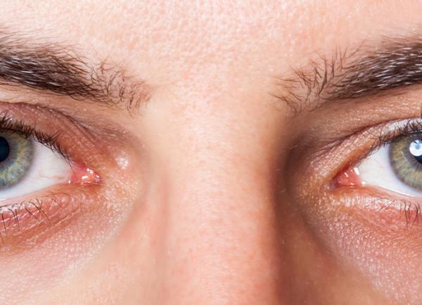
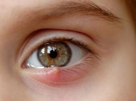
Blepharitis is chronic inflammation of the eyelid margins, typically due to bacterial colonization, meibomian gland dysfunction (MGD), or Demodex mite infestation. Cardinal features are eyelid itching or burning, crusting, redness, and fluctuating irritation that is often worse on awakening. Diagnosis is clinical at the slit lamp based on characteristic lid margin and lash findings. Management centers on eyelid hygiene; adjunctive topical or oral medications and in‑office procedures are used for flares, refractory disease, or specific subtypes.

Key Points

  • Blepharitis is a chronic, often relapsing eyelid margin inflammation driven by anterior factors (staphylococcal biofilm, seborrhea, Demodex) and/or posterior meibomian gland dysfunction.
  • Symptoms include burning, foreign‑body sensation, crusting, and eyelash debris; signs include lid margin erythema/telangiectasia, collarettes (cylindrical dandruff), scurf, capped glands, and turbid meibum.
  • Slit‑lamp examination is diagnostic; laboratory testing is rarely required except to confirm Demodex when uncertain or to evaluate atypical, refractory, or ulcerative presentations.
  • First‑line therapy is consistent eyelid hygiene (warm compresses, lid massage, and cleansers); add antibiotics, anti‑inflammatories, anti‑parasitics, or oral tetracyclines for targeted indications.
  • Demodex blepharitis is treated with lotilaner ophthalmic solution 0.25% (FDA‑approved) or, off‑label, tea tree oil–based regimens; MGD may benefit from thermal pulsation, manual expression, or intense pulsed light.
  • Complications include hordeolum, chalazion, trichiasis, keratoconjunctivitis, contact lens intolerance, and, rarely, corneal ulceration.

Anatomy and Physiology

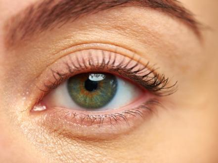
The eyelid margin contains the lash line (anterior lamella) with hair follicles, associated sebaceous glands (glands of Zeis), apocrine glands (glands of Moll), and the gray line bordering the posterior lamella that houses meibomian glands. Meibomian glands secrete meibum, the lipid layer of the tear film that retards evaporation and stabilizes the tear film. Eyelid margin keratinization, gland orifice patency, and balanced microbial flora maintain surface homeostasis.

Etiology

  • Anterior blepharitis
    • Staphylococcal biofilm and exotoxins (commonly coagulase‑negative staphylococci, Staphylococcus aureus)
    • Seborrheic dermatitis with increased scale at lash bases
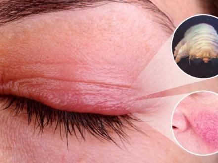
    • Demodex folliculorum infestation producing cylindrical collarettes
  • Posterior blepharitis (MGD)
    • Hyperkeratinization with orifice obstruction and altered meibum composition/flow
    • Rosacea‑associated meibomian gland changes
  • Contributing factors
    • Contact lens wear, ocular allergy, chronic topical medication use (preservatives), retinoids, androgen deficiency, dermatologic disease (seborrhea, psoriasis, atopy), and environmental desiccation

Pathophysiology

Biofilm and bacterial exotoxins at the anterior lid margin promote chronic inflammation and hypersensitivity, destabilize the tear film, and can incite marginal sterile keratitis. In MGD, ductal hyperkeratinization and increased meibum viscosity obstruct gland outflow, causing stasis, bacterial overgrowth, and lipase‑mediated alteration of meibum to pro‑inflammatory free fatty acids. Demodex mites and their antigens trigger follicular inflammation; their cylindrical dandruff (collarettes) trap bacteria and debris. These processes increase tear film osmolarity, reduce stability, and contribute to evaporative dry eye.

Epidemiology

Blepharitis is common in adults and increases with age. It frequently coexists with dry eye disease and rosacea and is a leading cause of ophthalmology and optometry visits in the United States [1],[2]. Demodex prevalence rises in older adults. Pediatric cases occur, often staphylococcal or associated with seborrhea or lice infestation.

Classification

  • By location
    • Anterior blepharitis: affects lash line and anterior lid margin (staphylococcal, seborrheic, Demodex)
    • Posterior blepharitis: meibomian gland dysfunction affecting posterior lid margin and tear film
    • Mixed: features of both
  • By specific cause
    • Staphylococcal
    • Seborrheic
    • Demodex
    • Ocular rosacea‑associated MGD

Symptoms and Signs

  • Common symptoms (all variants)
    • Burning, stinging, foreign‑body sensation, itching, photophobia, tearing, morning stickiness, fluctuating blurry vision (tear film instability), contact lens intolerance
  • Anterior staphylococcal
    • Symptoms: irritation, crusting on awakening
    • Signs: lid margin erythema, ulcerative crusts/scurf at lash bases, collarettes less prominent, lash breakage/madarosis in chronic cases
  • Seborrheic
    • Symptoms: greasy scales, less irritation
    • Signs: greasy soft scales along lashes, less erythema/ulceration, concomitant scalp/face seborrhea
  • Demodex blepharitis
    • Symptoms: itching disproportionate to exam, chronicity, poor response to standard lid hygiene
    • Signs: pathognomonic cylindrical dandruff (collarettes) at lash bases; visible mites on epilated lashes under light microscopy
  • Posterior (MGD/rosacea)
    • Symptoms: dryness, burning, blurred vision that clears after blinking
    • Signs: telangiectatic lid margin, orifice plugging/capping, toothpaste‑like turbid meibum on expression, frothy tear film, shortened tear breakup time

Complications

  • Hordeolum (external/internal) and chalazion recurrence
  • Trichiasis, poliosis, madarosis, lid margin notching
  • Conjunctivitis (bacterial or sterile) and keratoconjunctivitis
  • Marginal sterile keratitis, phlyctenules, superficial punctate keratopathy
  • Corneal ulceration, especially with contact lens wear or severe inflammation
  • Tear film instability with evaporative dry eye and contact lens intolerance

Diagnosis

Clinical evaluation

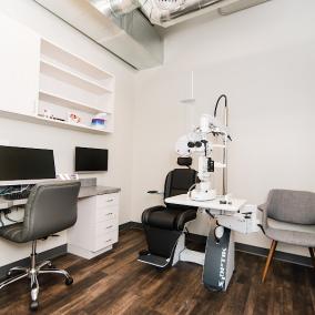
Diagnosis is clinical using slit‑lamp biomicroscopy:

  • Anterior: scurf, crusts, collarettes, lash misdirection/loss, ulceration
  • Posterior: capping of meibomian orifices, thickened/telangiectatic margin, turbid meibum on gentle expression, unstable tear film (short breakup time)
  • Demodex: cylindrical collarettes at lash bases; confirm by epilating lashes and visualizing mites

Assess for associated dermatologic disease (seborrhea, rosacea), medication history, contact lens wear, and prior episodes of hordeolum/chalazion.

Imaging and laboratory testing

  • Meibography can document gland dropout or truncation in MGD (adjunctive).
  • Lash sampling/microscopy confirms Demodex when diagnosis is uncertain or before targeted therapy.
  • Bacterial cultures are rarely needed; consider for severe ulcerative blepharitis, recurrent infections, immunocompromise, or atypical course.
  • Evaluate cornea for keratitis/ulcer if pain, photophobia, or decreased vision are present.

Differential diagnosis

Entity Distinguishing features
Dry eye disease (keratoconjunctivitis sicca) Burning/foreign‑body sensation; positive staining; reduced Schirmer; minimal lash debris; symptoms worse later in day
Allergic conjunctivitis Itching predominates; papillae; stringy discharge; seasonal or allergen exposure; chemosis
Bacterial conjunctivitis Mucopurulent discharge throughout day; conjunctival injection; minimal lash collarettes; self‑limited 7–10 days
Ocular rosacea without blepharitis Facial flushing, telangiectasias, phymatous changes; may have MGD overlap
Herpetic blepharitis Vesicular lid lesions, skin ulcerations, dendrites if cornea involved; segmental distribution
Pediculosis (Phthiriasis palpebrarum) Lice/nits on lashes; intense itching; blue‑gray maculae on skin
Contact dermatitis Periorbital erythema/edema, scaling; history of new cosmetics/drops
Preseptal/orbital cellulitis Tenderness, edema, fever; orbital signs (painful EOMs, proptosis, decreased vision) in orbital cellulitis—emergency
Molluscum contagiosum Umbilicated lid lesions with chronic follicular conjunctivitis
Trichiasis/entropion Misdirected lashes causing keratopathy; mechanical irritation predominates

Treatment

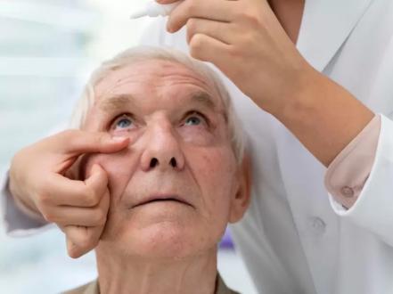
Chronic disease requires maintenance eyelid hygiene; intensify therapy during flares or for defined subtypes. Treat associated skin conditions (seborrhea, rosacea) and optimize contact lens hygiene.

Medical management

  • Lid hygiene (foundation of care) [1],[2]
    • Warm compresses: apply to closed lids 5–10 minutes, 1–2 times/day; followed by gentle lid massage to express meibum.
    • Lid cleansers: commercially prepared foam/wipes (eg, hypochlorous acid 0.01%–0.02% sprays or dilute non‑soap cleansers) to remove debris and biofilm. Avoid harsh detergents.
  • Topical antibiotics (anterior staphylococcal flares)
    • Erythromycin ophthalmic ointment 0.5%: thin ribbon to lid margins qhs (at bedtime) to bid for 2–8 weeks [1],[2].
    • Bacitracin ophthalmic ointment: thin ribbon to lid margins qhs to bid for 2–8 weeks [1].
    • Azithromycin 1% ophthalmic solution: off‑label for blepharitis/MGD; regimens vary (eg, 1 drop bid for 2 days, then daily for up to 2 weeks).
  • Anti‑inflammatory therapy (short courses for significant inflammation)
    • Loteprednol etabonate 0.5% or fluorometholone 0.1%: 1 drop qid for 1–2 weeks, then taper; monitor intraocular pressure and lens in prolonged/repeated use [1].
    • Cyclosporine ophthalmic emulsion 0.05% or lifitegrast 5%: off‑label adjunct in evaporative dry eye from MGD with ocular surface inflammation; onset over weeks.
  • Oral antibiotics (MGD/ocular rosacea or refractory disease)
    • Doxycycline: 50–100 mg PO bid for 2–4 weeks, then 50–100 mg daily (or 20–40 mg daily) for 1–3 months; counsel on photosensitivity, GI upset, and esophagitis; contraindicated in pregnancy and in children <8 years [1],[3].
    • Azithromycin: off‑label; example regimens include 500 mg on day 1 then 250 mg daily for 4 days, or 500 mg once weekly for 3 weeks.
  • Demodex‑directed therapy
    • Lotilaner ophthalmic solution 0.25% (Xdemvy): 1 drop in each eye bid for 6 weeks; FDA‑approved for Demodex blepharitis [4].
    • Tea tree oil/terpinen‑4‑ol–based lid hygiene: in‑office debridement and at‑home low‑concentration cleansers; may cause irritation; evidence quality variable [5].
    • Off‑label alternatives: topical ivermectin 1% cream applied carefully to lid margins nightly for several weeks; avoid ocular surface exposure.
  • Adjunctive dry eye care (as indicated)
    • Preservative‑free artificial tears qid or more frequently; nighttime lubricating ointments.
    • Environmental and behavioral modifications (humidification, screen breaks).

Note: Avoid chronic or repeated topical antibiotic‑steroid combinations unless closely supervised; minimize preservative exposure by using unit‑dose tears when possible.

Procedural and surgical management

  • In‑office lid debridement/microblepharoexfoliation to remove biofilm and collarettes (eg, device‑assisted “BlephEx”); repeat periodically in recalcitrant anterior blepharitis [2],[5].
  • Meibomian gland thermal pulsation (eg, LipiFlow), manual expression, or intense pulsed light (IPL) therapy for obstructive MGD; improves gland function in selected patients; multiple sessions may be needed; coverage varies [3],[5].
  • Meibomian gland probing for focal fibrotic obstruction (select cases).
  • Epilation of misdirected lashes for symptomatic trichiasis; definitive entropion/trichiasis repair if structural malposition exists.
  • Manage chalazia with intralesional corticosteroid injection or incision and curettage when conservative measures fail.

Special populations

  • Pediatric
    • Emphasize gentle lid hygiene; topical erythromycin or bacitracin ointment can be used for staphylococcal anterior disease.
    • Avoid tetracyclines in children <8 years; consider oral azithromycin off‑label in selected refractory MGD under pediatric/ophthalmology supervision.
    • Screen for lice (phthiriasis) and atopic dermatitis.
  • Pregnancy/lactation
    • Prefer non‑pharmacologic hygiene. Avoid tetracyclines (doxycycline, minocycline). Erythromycin ophthalmic is generally considered low risk. Discuss risks/benefits of any topical steroid; use the lowest effective dose and duration [6].
  • Immunocompromised/elderly
    • Lower threshold for culture in ulcerative cases; monitor closely for corneal involvement. Demodex prevalence is higher in older adults.

Prognosis

Blepharitis is typically chronic and relapsing. With consistent eyelid hygiene and targeted adjunctive therapy, most patients achieve symptom control and reduce flares. Relapses occur when maintenance lapses or in the presence of rosacea, Demodex, or environmental triggers. Visual prognosis is good; corneal complications are uncommon with appropriate care.

Prevention and Patient Counseling

  • Perform daily lid hygiene indefinitely even when asymptomatic; increase frequency during flares.
  • Use warm compresses and non‑irritating lid cleansers; replace eye makeup regularly and avoid during active flares.
  • Optimize contact lens wear: reduced wear time during flares, rub‑and‑rinse cleaning, avoid sleeping in lenses.
  • Manage dermatologic comorbidities (seborrhea, rosacea); minimize environmental desiccation (humidifier, screen breaks).
  • Avoid rubbing eyelids and harsh detergents on lids.

When to Seek Immediate Care

  • Sudden decrease in vision, severe eye pain, or light sensitivity
  • Redness spreading beyond the eyelids, fever, or feeling very ill
  • A white spot on the cornea, thick discharge, or inability to open the eye
  • Painful eye in a contact lens wearer
  • Swelling, redness, and pain with eye movement or bulging of the eye

References

  1. American Academy of Ophthalmology Cornea/External Disease Panel. Preferred Practice Pattern: Blepharitis. San Francisco, CA: AAO; 2018. https://www.aao.org/preferred-practice-pattern/blepharitis-ppp-2018
  2. EyeWiki. Blepharitis. American Academy of Ophthalmology. https://eyewiki.org/Blepharitis (accessed July 24, 2026).
  3. Geerling G, Tauber J, Baudouin C, et al. The international workshop on meibomian gland dysfunction: Report of the subcommittee on management and treatment. Invest Ophthalmol Vis Sci. 2011;52(4):2050-2064. doi:10.1167/iovs.10-6997g
  4. U.S. Food and Drug Administration. XDEMVY (lotilaner ophthalmic solution) 0.25% prescribing information. 2023. https://www.accessdata.fda.gov
  5. Lindsley K, Matsumura S, Hatef E, Akpek EK. Interventions for chronic blepharitis. Cochrane Database Syst Rev. 2012;(5):CD005556. doi:10.1002/14651858.CD005556.pub2
  6. Drugs and Lactation Database (LactMed). National Library of Medicine. https://www.ncbi.nlm.nih.gov/books/NBK501922/ (accessed July 24, 2026).
  7. NEI (National Eye Institute). Blepharitis. https://www.nei.nih.gov/learn-about-eye-health/eye-conditions-and-diseases/blepharitis (accessed July 24, 2026).
  8. StatPearls. Blepharitis. StatPearls Publishing; 2024. https://www.ncbi.nlm.nih.gov/books/NBK459305/

ICD‑10‑CM: H01.0 (Blepharitis)

Disclaimer: This article is for informational purposes and is not a substitute for professional medical advice, diagnosis, or treatment. Always consult a qualified eye care professional about your specific condition. If you have sudden vision loss, severe eye pain, or an eye injury, seek emergency care immediately.