Our eyes, the silent sentinels of our daily lives, often bear the brunt of hidden challenges. Glaucoma, a seemingly elusive term, encompasses a range of eye issues that, if left undetected, can lead to serious consequences for our vision. This eye-centric approach to understanding ocular well-being is crucial in a world where the complexities of vision care often remain underrated.
Much like computer vision syndrome, this eye issue stems from our modern lifestyles, where constant exposure to digital screens and other factors can lead to the occurrence of hidden inaccuracies within our eyes. Despite its prevalence, glaucoma often operates in the shadows, quietly affecting individuals without overt symptoms until the condition has advanced.
Imagine the scenario: you go about your daily routine, unaware that within your eyes, a subtle battle is taking place. This hidden adversary, if not addressed, could lead to irreversible vision impairment. Recognizing the triggers, signs, and preventive measures for this eye issue is important for maintaining long-term eye health.
The following sections describe its forms, causes, and care options. The sections below cover its signs, causes, and prevention.
Much like computer vision syndrome, this eye issue stems from our modern lifestyles, where constant exposure to digital screens and other factors can lead to the occurrence of hidden inaccuracies within our eyes. Despite its prevalence, glaucoma often operates in the shadows, quietly affecting individuals without overt symptoms until the condition has advanced.
Imagine the scenario: you go about your daily routine, unaware that within your eyes, a subtle battle is taking place. This hidden adversary, if not addressed, could lead to irreversible vision impairment. Recognizing the triggers, signs, and preventive measures for this eye issue is important for maintaining long-term eye health.
The following sections describe its forms, causes, and care options. The sections below cover its signs, causes, and prevention.
Glaucoma Causes: What Contributes to The Eye Issue
This eye issue, like a complex puzzle, has causes that are as intriguing as they are crucial to understand. Delving into the bedrock upon which this condition stands, we find that the factors contributing to its development are not solely reliant on one aspect but often a combination of various elements.
A key factor in this eye issue is raised intraocular pressure (IOP). Picture your eye as a delicate balance, where fluids continuously circulate to maintain optimal pressure. However, when this equilibrium is disrupted, and pressure within the eye rises, it becomes a potential trigger for vision complications.
Beyond just being paralyzed by high IOP, there are other hidden contributors to this eye issue. Genetics, for instance, plays a role in predisposing individuals to the condition. If you have family members grappling with vision challenges, the likelihood of encountering similar issues becomes a noteworthy consideration.
Environmental factors also emerge as key players in this eye issue's development. Prolonged exposure to certain substances or chronic conditions can act as catalysts, further complicating the eye's delicate balance. Much like pieces of a puzzle, these causes interconnect, shaping the narrative of how this eye issue manifests in different individuals.
Taken together, these glaucoma causes show that understanding the complex set of elements that lead to this eye issue is crucial.
A key factor in this eye issue is raised intraocular pressure (IOP). Picture your eye as a delicate balance, where fluids continuously circulate to maintain optimal pressure. However, when this equilibrium is disrupted, and pressure within the eye rises, it becomes a potential trigger for vision complications.
Beyond just being paralyzed by high IOP, there are other hidden contributors to this eye issue. Genetics, for instance, plays a role in predisposing individuals to the condition. If you have family members grappling with vision challenges, the likelihood of encountering similar issues becomes a noteworthy consideration.
Environmental factors also emerge as key players in this eye issue's development. Prolonged exposure to certain substances or chronic conditions can act as catalysts, further complicating the eye's delicate balance. Much like pieces of a puzzle, these causes interconnect, shaping the narrative of how this eye issue manifests in different individuals.
Taken together, these glaucoma causes show that understanding the complex set of elements that lead to this eye issue is crucial.
Types of Glaucoma
Now, it is time to arm yourselves with knowledge of condition types to be able to recognize them on time. Just as there are diverse landscapes in the world, this eye issue unfolds in various forms, each presenting unique challenges. By understanding the nuances of these different types, you empower yourself to decipher the subtle signals your eyes may be sending and take proactive steps toward preserving your vision.
Primary Open-Angle Type
One often overlooked eye issue is primary open-angle. Imagine a scenario where your eyes are akin to a finely tuned camera, capturing the world around you. However, this condition disrupts the delicate mechanism, quietly ushering in a gradual and unnoticed deterioration.
Primary open-angle, the silent architect of potential vision loss, is like a skilled infiltrator, creeping into our eyes without clamor or warning. Picture this: the drainage system of your eyes encounters a subtle blockage. This impediment, though inconspicuous, creates a slow but persistent increase in intraocular pressure, culminating in potential vision deterioration over time.
Primary open-angle, the silent architect of potential vision loss, is like a skilled infiltrator, creeping into our eyes without clamor or warning. Picture this: the drainage system of your eyes encounters a subtle blockage. This impediment, though inconspicuous, creates a slow but persistent increase in intraocular pressure, culminating in potential vision deterioration over time.
Primary Angle-Closure Glaucoma
Another eye issue is the primary angle closure condition that is beset by trouble with the potential to disrupt your visual harmony. Unlike its counterpart, this eye issue operates like a sudden storm, swiftly descending upon the ocular landscape.
Imagine your eye as a well-orchestrated performance, where the drainage system faces an unexpected blockage, causing pressure to build rapidly. Primary angle closure, like a hidden saboteur, thwarts the usual flow of fluids within the eye, creating a cascade of events that can prevail over optimal vision.
Here are the main characteristics of this eye issue. Vision can dim suddenly. Primary angle closure often manifests abruptly, causing a rapid onset of symptoms that demand immediate attention. These glaucoma symptoms, akin to warning signals, include intense eye pain, blurred vision, and halos around lights - a trifecta of indicators urging swift action.
Navigating the maze of primary angle closure involves understanding its swift onset and recognizing the unique signs that accompany it.
Here are the main characteristics of this eye issue. Vision can dim suddenly. Primary angle closure often manifests abruptly, causing a rapid onset of symptoms that demand immediate attention. These glaucoma symptoms, akin to warning signals, include intense eye pain, blurred vision, and halos around lights - a trifecta of indicators urging swift action.
Navigating the maze of primary angle closure involves understanding its swift onset and recognizing the unique signs that accompany it.
Secondary Glaucoma
In our exploration of eye health, we encounter yet another intricate facet - secondary vision complications. Unlike the swift storms of primary angle closure or the subtle erosion of primary open-angle, this eye issue, deemed as a byproduct of various underlying conditions, requires a closer examination.
Consider your eyes as sentinels, vigilant in their duty but susceptible to meager influences. Secondary vision complications, like a dormant force, lie in wait, ready to manifest when triggered by other health issues. Picture this scenario: an individual grappling with a chronic ailment, completely unaware of the potential implications for their eye health.
The meager presence of this eye issue often goes unnoticed until symptoms manifest. It can be intricately woven into the fabric of broader health concerns, making it imperative to recognize the interconnectedness of our well-being. The redemption lies in deciphering the underlying conditions that may give rise to secondary vision complications and addressing them holistically.
Consider your eyes as sentinels, vigilant in their duty but susceptible to meager influences. Secondary vision complications, like a dormant force, lie in wait, ready to manifest when triggered by other health issues. Picture this scenario: an individual grappling with a chronic ailment, completely unaware of the potential implications for their eye health.
The meager presence of this eye issue often goes unnoticed until symptoms manifest. It can be intricately woven into the fabric of broader health concerns, making it imperative to recognize the interconnectedness of our well-being. The redemption lies in deciphering the underlying conditions that may give rise to secondary vision complications and addressing them holistically.
Which Type of Glaucoma is the Most Serious?
In the landscape of vision complications, discerning the gravity of each concern is crucial. Amongst the various types, one stands out for its potential severity - primary angle closure. This eye issue, marked by its sudden onset and intense symptoms, poses an immediate threat to vision health. Picture this: an abrupt increase in intraocular pressure, causing rapid and severe symptoms such as intense eye pain and blurred vision. Unlike its counterparts, primary angle closure demands swift attention due to its potential to cause rapid and irreversible vision impairment.
Glaucoma Signs
Understanding the potential signals of this eye issue is paramount. These signals, known as glaucoma symptoms, often seem to be subtle whispers, easily dismissed until they snag attention with unwavering persistence.
Imagine this scenario: you've been handling adversity at work, spending long hours in front of screens. Instead of waiting for overt signs, it's crucial to tune into the nuances your eyes may be signaling. One common indicator is persistent eye pain, especially after prolonged screen time. The eyes, seemingly fatigued, may also prove to be more sensitive to light.
Among other signs, blurred vision can be another subtle warning. Moments of visual haziness, particularly after focusing on a task for an extended period, shouldn't be overlooked. Another red flag is the appearance of halos around lights, a phenomenon that may occur seemingly out of nowhere.
It's essential to recognize that these signals, though subtle, can be indicative of underlying issues with your eye health
Imagine this scenario: you've been handling adversity at work, spending long hours in front of screens. Instead of waiting for overt signs, it's crucial to tune into the nuances your eyes may be signaling. One common indicator is persistent eye pain, especially after prolonged screen time. The eyes, seemingly fatigued, may also prove to be more sensitive to light.
Among other signs, blurred vision can be another subtle warning. Moments of visual haziness, particularly after focusing on a task for an extended period, shouldn't be overlooked. Another red flag is the appearance of halos around lights, a phenomenon that may occur seemingly out of nowhere.
It's essential to recognize that these signals, though subtle, can be indicative of underlying issues with your eye health
What is The Best Way to Diagnose Glaucoma?
Understanding how to accurately diagnose this eye issue is paramount for preserving your eye health. The process involves a careful unraveling of subtle signals and a reliance on advanced diagnostic methods that delve into the intricate workings of the eyes.
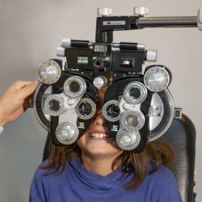
Imagine that you've noticed persistent eye symptoms, prompting the need for an examination. Instead of relying solely on apparent signs, healthcare professionals employ a variety of tests to decipher the health of your eyes. Among these, the visual field test stands out as one of the most accurate tests for this eye issue. Picture it as a detailed map of your vision, where any anomalies can be precisely identified, aiding in early detection.
In addition to the visual field test, another vital tool in the diagnostic arsenal is tonometry. This method measures intraocular pressure, a key factor in determining the presence of this eye issue. While tonometry might seem like a routine eye check, it plays a crucial role in assessing the risk of potential vision loss.
Navigating the path to an accurate diagnosis involves a multi-faceted approach, combining patient history, comprehensive eye examinations, and advanced tests.
Imagine that you've noticed persistent eye symptoms, prompting the need for an examination. Instead of relying solely on apparent signs, healthcare professionals employ a variety of tests to decipher the health of your eyes. Among these, the visual field test stands out as one of the most accurate tests for this eye issue. Picture it as a detailed map of your vision, where any anomalies can be precisely identified, aiding in early detection.
In addition to the visual field test, another vital tool in the diagnostic arsenal is tonometry. This method measures intraocular pressure, a key factor in determining the presence of this eye issue. While tonometry might seem like a routine eye check, it plays a crucial role in assessing the risk of potential vision loss.
Navigating the path to an accurate diagnosis involves a multi-faceted approach, combining patient history, comprehensive eye examinations, and advanced tests.
What's the Treatment for Glaucoma?
- Medications. When it comes to managing this eye issue, a well-rounded treatment plan often involves medications as a cornerstone. Imagine these medications as skilled navigators, synchronizing treatment efforts to maintain optimal eye health. The first category of medications geared toward managing this eye issue includes eye drops. These specialized drops work in different ways - some decrease intraocular pressure by enhancing fluid drainage, while others reduce the production of aqueous humor, the fluid within the eye. It's like a meticulous dance within your eyes, where these drops play a crucial role in maintaining the delicate balance. An affected individual has to diligently apply prescribed eye drops as part of their treatment regimen. The aim is not just to alleviate symptoms but to proactively address the underlying factors contributing to this eye condition. It urges actions beyond symptom management, focusing on the preservation of vision over the long term. Medications, including oral medications, can also be part of the arsenal in managing this eye issue. These may be prescribed when eye drops alone prove insufficient. As we delve into the realm of medication-based treatments, it becomes evident that these pharmacological interventions play a pivotal role in curbing the progression of this eye issue, providing individuals with a proactive means of maintaining optimal vision health.
- Laser Therapy. In the arsenal of treatments for this eye issue, laser therapy emerges as a beacon of precision. Imagine a scenario where targeted beams of light are strategically employed to enhance drainage within the eye, reducing intraocular pressure. This innovative approach showcases the strides made in providing effective and less invasive solutions for managing this eye issue. Individuals opting for laser therapy often experience a swift and relatively painless procedure, emphasizing the evolving landscape of treatments tailored to preserve optimal vision health.
- Trabeculectomy. When this eye issue demands more decisive measures, surgical interventions like trabeculectomy come into play. Think of it as a meticulous architectural endeavor within the eye, aimed at creating a drainage pathway to alleviate intraocular pressure. Trabeculectomy is like crafting an escape route for excess fluid, addressing the root cause of elevated pressure within the eye. A surgeon delicately creates a tiny channel, allowing fluid to flow out, akin to unclogging a blocked drain. This surgical procedure showcases the delicate precision required in managing this eye issue. For those seeking a less invasive option, selective laser trabeculoplasty (SLT) emerges as a modern marvel. Unlike traditional surgical interventions, SLT employs laser technology to enhance drainage, reducing intraocular pressure without the need for incisions. It's like a technological symphony, where precision meets innovation to tackle this eye issue effectively. Consider opting for SLT to undergo a relatively quick and painless procedure. This advanced treatment emphasizes targeted precision, reducing the risk of complications associated with traditional surgical approaches. As we explore the surgical landscape in managing this eye issue, trabeculectomy and SLT showcase the strides made in providing individuals with effective and tailored solutions.
Preventing Glaucoma
The journey of visual wellness must comprise understanding and integrating strategies for preventing this eye issue, and it becomes a crucial inroad to maintaining optimal eye health. While it's true that certain factors contributing to glaucoma may be beyond our control, there are proactive steps that can be taken to free up the path to prevention.
One key leap forward in preventing this eye issue is regular eye examinations. These balanced assessments serve as a re-framing tool, allowing healthcare professionals to detect potential issues early on. Picture it as a pause and refresh moment for your eyes, providing a comprehensive overview of their health.
For those at higher risk due to family history or other factors, specialized examinations may be necessary. Think of this as a targeted intervention, where healthcare professionals can identify potential red flags and tailor prevention strategies accordingly.
In the realm of visual wellness, understanding and implementing these strategies become a proactive endeavor. It's about more than just preserving your eyesight; it's about creating a foundation for visual health that fosters a fast recovery from potential threats.
Remember, glaucoma prevention involves more than just addressing symptoms - it's about cultivating habits that safeguard your vision for the long term.
One key leap forward in preventing this eye issue is regular eye examinations. These balanced assessments serve as a re-framing tool, allowing healthcare professionals to detect potential issues early on. Picture it as a pause and refresh moment for your eyes, providing a comprehensive overview of their health.
For those at higher risk due to family history or other factors, specialized examinations may be necessary. Think of this as a targeted intervention, where healthcare professionals can identify potential red flags and tailor prevention strategies accordingly.
In the realm of visual wellness, understanding and implementing these strategies become a proactive endeavor. It's about more than just preserving your eyesight; it's about creating a foundation for visual health that fosters a fast recovery from potential threats.
Remember, glaucoma prevention involves more than just addressing symptoms - it's about cultivating habits that safeguard your vision for the long term.