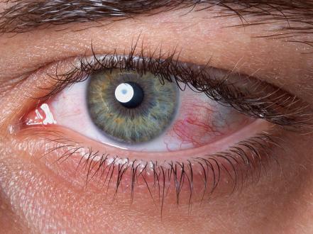
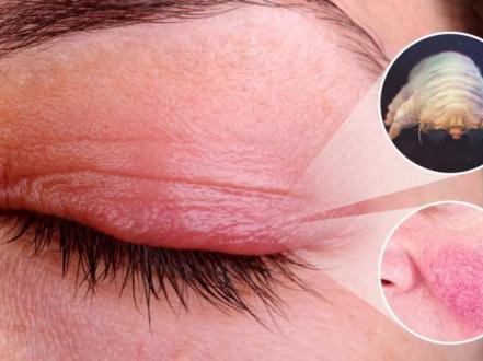
Keratitis is inflammation of the cornea, the transparent anterior surface of the eye. Causes include infection (bacteria, herpesviruses, fungi, Acanthamoeba) and noninfectious insults (contact lens overwear, exposure, trauma, ultraviolet light, autoimmune disease). Pain, photophobia, tearing, red eye, and reduced vision are typical. Diagnosis is clinical with slit-lamp biomicroscopy; corneal scrapings and targeted tests are used for sight-threatening or atypical cases. Prompt, etiology-specific therapy (topical antimicrobials, antivirals, anti-inflammatories, or systemic immunosuppression) prevents scarring, thinning, and perforation.

Key Points

  • Keratitis may be infectious or noninfectious; history of contact lens wear, trauma with organic matter, or facial/shingles rash guides etiology.
  • Central, large, or deep ulcers require urgent culture-guided therapy; do not start corticosteroids when fungal or Acanthamoeba keratitis is suspected.
  • Fluoroquinolone monotherapy treats many mild bacterial ulcers; severe ulcers need fortified antibiotics and close follow-up.
  • Herpes simplex epithelial keratitis is treated with topical or oral antivirals; avoid topical steroids until the epithelium heals.
  • Fungal keratitis requires natamycin 5% for filamentous organisms; Acanthamoeba keratitis needs biguanide-based combination therapy over months.
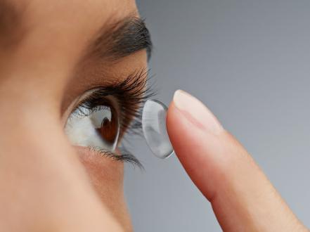
  • Contact lens hygiene lapses are a major, preventable risk; discontinue lens wear with any painful red eye.

Anatomy and Physiology

The cornea comprises five principal layers: epithelium (barrier with rapid turnover), Bowman layer (acellular), stroma (collagen lamellae, avascular), Descemet membrane (basement membrane of endothelium), and endothelium (pumps to maintain deturgescence). Dense trigeminal innervation mediates pain and blink reflex. The tear film provides antimicrobial peptides and immunoglobulins; eyelids distribute and replenish tears. Any disruption of epithelial integrity or tear film predisposes to keratitis.

Etiology

  • Infectious
    • Bacterial: Staphylococcus aureus, coagulase-negative staphylococci, Streptococcus pneumoniae, Pseudomonas aeruginosa (notably in contact lens wearers), Gram-negative rods, Nocardia.
    • Viral: Herpes simplex virus type 1 (HSV-1), varicella-zoster virus (VZV; herpes zoster ophthalmicus).
    • Fungal: Filamentous (Fusarium, Aspergillus) and yeasts (Candida), often after plant matter trauma or chronic surface disease.
    • Protozoal: Acanthamoeba, associated with contact lens exposure to water (tap, swimming, hot tubs) and soil.
  • Noninfectious
    • Mechanical/toxic: Contact lens overwear, foreign body, topical anesthetic abuse, preservatives.
    • Exposure and neurotrophic keratopathy: Lagophthalmos, facial nerve palsy, reduced corneal sensation from diabetes, prior HSV, surgery.
    • Immune-mediated: Staphylococcal marginal keratitis (hypersensitivity), peripheral ulcerative keratitis (PUK) in rheumatoid arthritis, granulomatosis with polyangiitis, Mooren ulcer.
    • Radiation: Photokeratitis from ultraviolet (UV) exposure (welding arcs, high-altitude snow, tanning beds).
    • Chemical: Alkali/acid burns.

Pathophysiology

Epithelial compromise permits microbial adherence and stromal invasion. Bacterial keratitis triggers neutrophil influx, collagenolysis, and tissue necrosis; Pseudomonas elaborates proteases and biofilm that rapidly liquefy stroma. HSV replicates in epithelium causing dendritic ulceration; immune responses to viral antigens drive stromal keratitis. Fungal filaments extend within stroma with slow, indolent inflammation; yeasts colonize compromised epithelium. Acanthamoeba trophozoites invade nerves and stroma, provoking perineural inflammation and disproportionate pain. In immune keratitides, circulating immune complexes and local cytokines induce peripheral stromal melt.

Epidemiology

Microbial keratitis risk is highest in contact lens wearers, particularly with overnight wear and poor hygiene. The US Centers for Disease Control and Prevention estimates about 1 in 500 contact lens users develop a serious eye infection each year, many preventable through proper care [1]. HSV eye disease is common and recurrent; antiviral prophylaxis reduces recurrence risk [2]. Fungal keratitis in the United States is less frequent than bacterial, but more prevalent in warm, humid regions and after vegetative trauma [3]. Acanthamoeba keratitis remains rare but severe; most US cases occur in soft contact lens users with water exposure [4].

Classification

  • By cause: infectious (bacterial, viral, fungal, protozoal) vs noninfectious (exposure, neurotrophic, immune, toxic, UV).
  • By anatomic depth: superficial punctate keratitis, epithelial ulcer, stromal/interstitial keratitis, endothelial/keratic precipitates.
  • By location: central (visual axis), paracentral, peripheral (limbus-adjacent).
  • By severity: size (>2 mm), depth (>50% stromal thinning), presence of hypopyon, rapid progression.
  • By context: contact lens–related, trauma-related, postoperative, immunocompromised host.

Symptoms and Signs

Bacterial keratitis

  • Symptoms: Acute pain, photophobia, foreign body sensation, mucopurulent discharge, decreased vision; rapid onset, especially in contact lens wearers.
  • Signs: Focal stromal infiltrate with overlying epithelial defect (fluorescein-positive), dense suppuration, surrounding edema, anterior chamber cells/flare, possible hypopyon. Pseudomonas may produce a gray-green, rapidly spreading ulcer with stromal melt.

Herpes simplex keratitis

  • Symptoms: Pain, photophobia, tearing; recurrent episodes; reduced corneal sensation possible.
  • Signs: Branching dendritic epithelial ulcer with terminal bulbs that stain with fluorescein; geographic ulcers if steroid-exposed; disciform stromal edema/keratitis in immune-mediated phases; keratic precipitates; decreased corneal sensitivity.

Herpes zoster ophthalmicus keratitis

  • Symptoms: Dermatomal forehead/eyelid pain and vesicular rash (V1), ocular pain and photophobia.
  • Signs: Pseudodendrites (staining pattern different from HSV), nummular anterior stromal infiltrates, epithelial defects; concurrent anterior uveitis.

Fungal keratitis

  • Symptoms: Subacute pain, photophobia, foreign body sensation following plant matter trauma or chronic ocular surface disease.
  • Signs: Feathery-edged stromal infiltrate, dry surface, satellite lesions, possible endothelial plaque or hypopyon; slower progression than bacterial.

Acanthamoeba keratitis

  • Symptoms: Severe pain out of proportion to findings, photophobia; history of soft contact lens wear and water exposure.
  • Signs: Punctate keratitis progressing to ring-shaped stromal infiltrate, perineural infiltrates (radial keratoneuritis), epithelial defects variable; often misdiagnosed early.

Noninfectious keratitis

  • Photokeratitis: Delayed (6–12 hours) severe photophobia, tearing, blepharospasm after UV exposure; diffuse punctate epithelial erosions.
  • Exposure/neurotrophic keratopathy: Dulling of epithelium, inferior punctate staining, persistent epithelial defect with rolled edges, reduced corneal sensitivity.
  • Immune marginal keratitis: Peripheral arcuate subepithelial infiltrates adjacent to limbus, clear corneal zone between infiltrate and limbus; associated blepharitis.
  • PUK/Mooren: Crescentic peripheral thinning with epithelial defect and stromal melt; adjacent conjunctival injection.

Complications

  • Corneal scarring and vascularization causing permanent visual loss.
  • Corneal thinning, descemetocele, and perforation; anterior synechiae.
  • Endophthalmitis from bacterial or fungal extension.
  • Secondary glaucoma; cataract (often steroid-related).
  • Irregular astigmatism and anisometropia; amblyopia risk in children.

Diagnosis

Clinical evaluation

  • History: Contact lens wear (overnight use, water exposure, solutions), trauma (particularly vegetative), prior herpes eye disease or shingles, topical medication/toxin exposure, systemic autoimmune disease, immunosuppression.
  • Examination: Visual acuity; external inspection; slit-lamp evaluation with fluorescein staining to define epithelial defect; assess infiltrate size, depth, location; anterior chamber reaction/hypopyon; intraocular pressure (avoid in thin corneas).
  • Corneal sensitivity testing if HSV/neurotrophic disease suspected.
  • Urgent ophthalmology evaluation for any central, large (>2 mm), deep, or rapidly progressive ulcer.

Imaging and laboratory testing

  • Corneal scrapings for Gram stain and culture (blood/chocolate agar) for bacterial ulcers that are central, large, deep, atypical, or unresponsive; add Sabouraud dextrose agar for fungi; non-nutrient agar with E. coli overlay for Acanthamoeba [5].
  • PCR from corneal/tear samples for HSV/VZV when clinical diagnosis is uncertain [6].
  • Potassium hydroxide (KOH) wet mount or calcofluor white for fungi; Giemsa for Acanthamoeba cysts/trophozoites.
  • In vivo confocal microscopy to visualize Acanthamoeba cysts or fungal hyphae when available.
  • Anterior segment OCT to gauge depth; corneal topography post-resolution for irregular astigmatism.
  • Autoimmune workup (RF, anti-CCP, ANCA) in suspected PUK with rheumatology consultation.

Differential diagnosis

Entity Distinguishing features
Bacterial conjunctivitis Conjunctival papillae and diffuse injection, mucopurulent discharge, minimal photophobia, no focal corneal infiltrate; see bacterial conjunctivitis.
Corneal abrasion Fluorescein-positive epithelial defect without stromal infiltrate; traumatic history; rapid healing in 24–72 hours.
Episcleritis Sectoral redness, mild tenderness, blanches with phenylephrine, no photophobia or corneal staining.
Scleritis Severe, boring pain, deep violaceous vessels that do not blanch, often systemic autoimmune disease.
Anterior uveitis (iritis) Photophobia and ciliary flush, cells/flare, keratic precipitates; no epithelial defect or focal infiltrate.
Acute angle-closure glaucoma Severe pain, headache, halos, mid-dilated fixed pupil, corneal edema, markedly elevated IOP.
Dry eye disease Chronic burning/grittiness, punctate staining without focal infiltrate; worse with screen time; see dry eye syndrome blog.
Chalazion/blepharitis-related Eyelid margin disease with collarettes; sterile marginal infiltrates separated from limbus by clear zone; see chalazion.

Treatment

Medical management

  • General measures
    • Discontinue contact lens wear immediately.
    • Cycloplegic for pain/photophobia: cyclopentolate 1% tid or homatropine 5% bid–tid.
    • Analgesia (PO NSAIDs or acetaminophen); avoid topical anesthetics outside the clinic.
  • Bacterial keratitis
    • Small, non-severe ulcers: topical fluoroquinolone monotherapy off-label (e.g., moxifloxacin 0.5%, gatifloxacin 0.5%, besifloxacin 0.6%, ciprofloxacin 0.3%, or ofloxacin 0.3%) 1 drop q1–2h while awake for 24–48 hours, then taper per response [5]. Avoid nighttime gaps early for progressive ulcers.
    • Severe, central, deep, or sight-threatening ulcers: fortified antibiotics (compounded) hourly around-the-clock initially—tobramycin 14 mg/mL alternating with cefazolin 50 mg/mL, or vancomycin 25–50 mg/mL if MRSA risk—then taper with clinical improvement [5].
    • Adjunct corticosteroids: may be started 24–48 hours after effective antibiotics in bacterial ulcers without suspicion for Nocardia or fungus; typical prednisolone acetate 1% qid then taper; based on SCUT outcomes [7]. Avoid steroids if etiology uncertain.
  • Herpes simplex keratitis
    • Epithelial disease: topical ganciclovir 0.15% gel 5×/day until healing, then tid for 7 days (FDA-approved), or oral acyclovir 400 mg PO five times daily 7–10 days or valacyclovir 500 mg PO tid 7–10 days [6]. Avoid topical steroids until epithelial healing.
    • Stromal/disciform disease: topical corticosteroid (e.g., prednisolone acetate 1% qid) WITH concurrent antiviral cover (topical or oral) and close follow-up [6].
    • Prophylaxis to reduce recurrences: acyclovir 400 mg PO bid for 12 months (HEDS) [2]; valacyclovir 500 mg PO daily is a common alternative.
  • Herpes zoster ophthalmicus
    • Start systemic antiviral within 72 hours: valacyclovir 1,000 mg PO tid or acyclovir 800 mg PO five times daily for 7 days; add topical steroids for keratitis/uveitis under ophthalmologist supervision [6].
  • Fungal keratitis
    • Filamentous fungi: natamycin 5% suspension 1 drop hourly while awake for several days, then taper slowly over weeks as infiltrate resolves (FDA-approved) [3].
    • Yeasts: amphotericin B 0.15% (compounded) or voriconazole 1% (compounded) topical hourly initially; consider oral voriconazole 200 mg PO bid for deep/large ulcers or endothelial involvement [3]. Avoid topical steroids.
  • Acanthamoeba keratitis
    • Combination topical biguanides (polyhexamethylene biguanide 0.02% and/or chlorhexidine 0.02%) hourly day and night initially, tapered over months; add diamidines (e.g., propamidine or hexamidine) if available; therapy is off-label in the US and prolonged [4].
    • Oral miltefosine has been used off-label as adjunct in recalcitrant cases.
    • Pain control, judicious debridement; delay corticosteroids early; consider later for scleritis under specialist care.
  • Noninfectious keratitis
    • Photokeratitis: lubricants, cycloplegic, oral analgesics; prophylactic topical antibiotic ointment (e.g., erythromycin) qid for 2–3 days; heals in 24–72 hours.
    • Exposure/neurotrophic keratopathy: preservative-free artificial tears qid–hourly, gel/ointment at night; moisture shields/taping; consider amniotic membrane or temporary tarsorrhaphy if persistent epithelial defect. Cenegermin 0.002% (Oxervate) 1 drop q6h for 8 weeks is FDA-approved for neurotrophic keratitis [8].
    • Immune marginal keratitis: lid hygiene, warm compresses, topical antibiotic if blepharitis; short course of low-dose topical corticosteroid for infiltrates once infection is excluded.
    • PUK: urgent systemic immunosuppression in collaboration with rheumatology (high-dose systemic corticosteroids; steroid-sparing agents). Doxycycline 100 mg PO bid and vitamin C 1 g/day are commonly used adjuncts to reduce corneal melt.

Procedural and surgical management

  • Corneal debridement to reduce microbial load and improve drug penetration (especially HSV and Acanthamoeba).
  • Corneal glue (cyanoacrylate) with bandage contact lens for small perforations (<2 mm); anterior chamber reformation if needed.
  • Amniotic membrane transplantation for persistent epithelial defects or melts (CPT 65778).
  • Therapeutic keratoplasty (deep anterior lamellar or penetrating) for nonresolving infection, descemetocele, or perforation (CPT 65730).
  • Corneal scraping for smear/culture (CPT 65430).
  • Intrastromal/intracameral antifungal injections for recalcitrant fungal keratitis under specialist protocols.
  • Corneal cross-linking as adjuvant for infectious keratitis remains investigational in the US (PACK-CXL).

Special populations

  • Pediatric: Higher amblyopia risk after scarring; urgent management and optical rehabilitation. HSV keratitis may mimic abrasions; consider prophylaxis for recurrent disease per HEDS principles.
  • Pregnancy/lactation: Oral acyclovir/valacyclovir are generally considered compatible; avoid doxycycline; limit topical medications to necessary agents; coordinate with obstetrics [6].
  • Immunocompromised: Higher risk of severe viral and fungal keratitis; lower threshold for culture, systemic therapy, and admission.
  • Elderly: Increased exposure and neurotrophic keratopathy due to eyelid laxity and reduced corneal sensitivity.

Prognosis

With prompt, appropriate therapy, many bacterial and herpetic ulcers heal with minimal scarring if extrafoveal. Central, large, or fungal/Acanthamoeba infections carry higher risks of scarring, thinning, or perforation and may require surgery. Recurrent HSV/VZV can lead to cumulative stromal damage and neurotrophic keratopathy without prophylaxis [2][6]. Visual outcome depends on location, depth, timely diagnosis, and organism virulence.

Prevention and Patient Counseling

  • Contact lens hygiene: no overnight wear unless approved; never expose lenses or cases to tap water; rub-and-rinse cleaning; replace cases every 3 months; discontinue lens wear with any painful red eye; do not “top off” solutions [1].
  • Eye protection: wear UV-blocking eyewear for welding, snow sports, and tanning devices to prevent photokeratitis.
  • Eyelid and surface care: manage blepharitis and meibomian gland dysfunction; treat dry eye disease to maintain an intact epithelium.
  • Systemic disease control: coordinate care for autoimmune disease to prevent PUK.
  • Avoid topical anesthetic abuse and nonsterile home remedies.

When to Seek Immediate Care

  • Sudden eye pain, light sensitivity, or vision loss.
  • A new white/gray spot on the cornea.
  • A painful red eye if you wear contact lenses.
  • Chemical splash or high-velocity eye injury.
  • Shingles rash on the forehead/eyelid with eye symptoms.
  • Thick discharge, fever, or symptoms that worsen despite treatment.

References

  1. Centers for Disease Control and Prevention (CDC). Contact Lens–Related Eye Infections and Keratitis: Fast Facts. 2023. https://www.cdc.gov/contactlenses/fast-facts.html
  2. The Herpetic Eye Disease Study Group. Acyclovir for the prevention of recurrent herpes simplex virus eye disease. N Engl J Med. 1998;339(5):300-306. doi:10.1056/NEJM199807303390503
  3. American Academy of Ophthalmology. Fungal Keratitis. EyeWiki. Updated 2024. https://eyewiki.org/Fungal_Keratitis
  4. American Academy of Ophthalmology. Acanthamoeba Keratitis. EyeWiki. Updated 2024. https://eyewiki.org/Acanthamoeba_Keratitis
  5. American Academy of Ophthalmology Preferred Practice Pattern. Bacterial Keratitis PPP. 2023. https://www.aao.org/preferred-practice-pattern/bacterial-keratitis-ppp
  6. American Academy of Ophthalmology Preferred Practice Pattern. Herpes Simplex Virus Keratitis PPP. 2023. https://www.aao.org/preferred-practice-pattern/herpes-simplex-virus-keratitis-disease-ppp
  7. Srinivasan M, Mascarenhas J, Rajaraman R, et al. The Steroids for Corneal Ulcers Trial (SCUT): results of a randomized, placebo-controlled trial. Arch Ophthalmol. 2012;130(2):143-150.
  8. DailyMed. Oxervate (cenegermin-bkbj) ophthalmic solution 0.002% prescribing information. 2018. https://dailymed.nlm.nih.gov/dailymed/drugInfo.cfm?setid=2b93e3b8-0e00-4a87-bb30-3a7ba1a41585

Disclaimer: This article is for informational purposes and is not a substitute for professional medical advice, diagnosis, or treatment. Always consult a qualified eye care professional about your specific condition. If you have sudden vision loss, severe eye pain, or an eye injury, seek emergency care immediately.