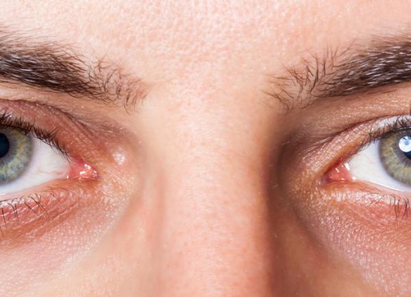
Conjunctivitis is inflammation of the bulbar and palpebral conjunctiva—the transparent mucous membrane covering the sclera and inner eyelids—caused most often by viruses, bacteria, or allergic reactions. Cardinal features are diffuse eye redness, discharge, burning or gritty irritation, tearing, and eyelid sticking/crusting. Diagnosis is clinical; routine laboratory testing is rarely needed. Management targets the cause: supportive care for viral disease, topical antihistamine/mast cell stabilizers for allergic disease, and topical or systemic antimicrobials when bacterial or sexually transmitted infections are suspected.
Key Points
- Most adult cases are viral (adenovirus) and self-limited; most pediatric cases are bacterial. Avoid routine antibiotics unless bacterial infection is likely or risk factors are present.
- Red eye with severe pain, photophobia, or vision decrease is not typical; evaluate urgently for keratitis, uveitis, or acute glaucoma.
- Contact lens wearers with a painful red eye require prompt evaluation for bacterial keratitis (including Pseudomonas); stop lens wear immediately.
- Do not use topical corticosteroids in suspected infectious conjunctivitis unless directed by an ophthalmologist (risk of masking or worsening herpes simplex or adenoviral keratitis).
- Neonatal conjunctivitis and hyperacute purulent discharge in any age suggest gonococcal or chlamydial infection and require urgent systemic therapy and specialist care.
- Hand hygiene, avoiding eye rubbing, and not sharing towels or cosmetics reduce transmission; no contact lens use until full resolution.
Anatomy and Physiology
The conjunctiva is a nonkeratinized stratified columnar epithelium with goblet cells overlying a vascular stroma. It lines the posterior eyelids (palpebral conjunctiva) and reflects onto the globe to cover the anterior sclera (bulbar conjunctiva). It forms the tear film’s mucin layer, participates in ocular surface immunity (lymphoid tissue, IgA), and permits smooth eyelid-globe movement.
Etiology
- Infectious
- Viral: adenoviruses (epidemic keratoconjunctivitis, pharyngoconjunctival fever); herpes simplex virus (HSV); varicella-zoster virus (VZV).
- Bacterial: Staphylococcus aureus, Streptococcus pneumoniae, Haemophilus influenzae, Moraxella spp.; Neisseria gonorrhoeae (hyperacute).
- Chlamydial: Chlamydia trachomatis (adult inclusion conjunctivitis; neonatal ophthalmia).
- Noninfectious
- Allergic: seasonal/perennial allergic conjunctivitis; vernal keratoconjunctivitis (VKC); atopic keratoconjunctivitis (AKC).
- Toxic/irritative: preservatives, topical glaucoma drugs (e.g., brimonidine), vasoconstrictor overuse (rebound), chemical or UV exposure.
- Mechanical/other: contact lens overwear, floppy eyelid syndrome, ocular surface disease (dry eye), blepharitis; autoimmune/rosacea-associated ocular surface inflammation.
Pathophysiology
- Viral: adenoviral replication triggers follicular conjunctivitis with preauricular lymphadenopathy; corneal epithelial involvement may lead to subepithelial infiltrates and photophobia (epidemic keratoconjunctivitis).
- Bacterial: pathogen adherence and biofilm on conjunctival/corneal epithelium produce purulent discharge and papillary reaction; N. gonorrhoeae can invade intact epithelium causing rapid keratitis and perforation.
- Chlamydial: intracellular replication in conjunctival epithelium causes chronic follicular conjunctivitis; in neonates, transmission occurs during delivery.
- Allergic: IgE-mediated mast cell degranulation with histamine release leads to itching, chemosis, tearing; VKC/AKC involve chronic Th2-mediated inflammation with risk of corneal damage.
- Toxic/irritative: direct epithelial toxicity and inflammatory cascade; chronic exposure causes medicamentosa.
Epidemiology
- Conjunctivitis is among the most common eye complaints in primary and urgent care. In adults, viral etiologies predominate; in children, bacterial causes are more common [1,2].
- Adenovirus is the leading viral cause; outbreaks occur in community and healthcare settings [3].
- Contact lens wear increases risk of bacterial conjunctivitis and keratitis (including Pseudomonas) [1].
- Neonatal conjunctivitis incidence is low in the US due to prophylaxis but remains critical because of gonococcal/chlamydial risk [4,5].
- ICD-10-CM: H10.- (e.g., H10.9 unspecified conjunctivitis; see subcodes by type and laterality).
Classification
- Infectious: viral, acute bacterial, hyperacute gonococcal, chlamydial (adult inclusion), neonatal ophthalmia (gonococcal, chlamydial).
- Noninfectious: allergic (seasonal/perennial), VKC, AKC; toxic/medicamentosa; irritative (chemical/UV); mechanical/contact lens-related; autoimmune-associated conjunctivitides.
Symptoms and Signs
Viral conjunctivitis (adenoviral)
- Watery or serous discharge, burning or gritty sensation, mild photophobia, bilateral involvement often sequential, preauricular lymphadenopathy, follicles on tarsal conjunctiva. Recent upper respiratory infection or sick contacts common. Pseudomembranes and corneal subepithelial infiltrates may develop in epidemic keratoconjunctivitis (EKC).
Bacterial conjunctivitis
- Mucopurulent discharge causing eyelid mattering (“glued” lids), conjunctival hyperemia, foreign-body sensation; typically starts in one eye and may spread. Papillary reaction on tarsal conjunctiva. Contact lens wear raises concern for Pseudomonas and keratitis.
Hyperacute gonococcal conjunctivitis
- Rapid onset, copious purulent discharge, marked chemosis and eyelid swelling, pain, and tender preauricular nodes; corneal involvement can progress quickly to ulceration/perforation.
Chlamydial (adult inclusion) conjunctivitis
- Chronic (>2–3 weeks) unilateral or bilateral redness with mucopurulent discharge, follicles, and preauricular nodes; may have concurrent genital symptoms or exposure.
Allergic conjunctivitis (seasonal/perennial)
- Prominent itching, tearing, chemosis, stringy/ropelike mucus, bilateral symptoms, papillae; eyelid dermatitis may coexist. VKC/AKC show severe itching, photophobia, large “cobblestone” papillae, shields ulcers risk.
Toxic/irritative/medicamentosa
- Burning, redness, tearing without significant discharge; history of new drops, cosmetics, or chemical exposure. Follicular reaction common with brimonidine or preserved drops.
Neonatal conjunctivitis (ophthalmia neonatorum)
- Onset and features vary by pathogen: 2–5 days (gonococcal—copious purulence, chemosis); 5–14 days (chlamydial—mucopurulent, follicles uncommon in neonates); within 24 hours suggests chemical irritation.
Complications
- Keratitis and corneal ulceration (notably with N. gonorrhoeae and contact lens-associated infections).
- Subepithelial corneal infiltrates after adenovirus with photophobia and decreased acuity.
- Membranes/pseudomembranes with conjunctival scarring, symblepharon (severe EKC, cicatrizing disease).
- Chronic conjunctival changes and corneal involvement in VKC/AKC.
- Secondary dry eye symptoms.
- Steroid complications if misused: elevated intraocular pressure, cataract, worsened herpetic disease.
Diagnosis
Clinical evaluation
- History: laterality, discharge character, itching (suggests allergy), contact lens wear, systemic symptoms, sexual history, neonatal risk factors, medication exposures.
- Examination: visual acuity; pattern of redness (diffuse vs sectoral); tarsal follicles vs papillae; preauricular lymph nodes; corneal assessment with fluorescein staining; anterior chamber reaction to exclude uveitis.
- Red-flag features (pain, photophobia, decreased vision, corneal staining, ciliary flush) prompt urgent ophthalmology referral.
Imaging and laboratory testing
- Routine cultures or PCR are not indicated for uncomplicated cases.
- Obtain conjunctival swab for Gram stain/culture or NAAT when: hyperacute purulence; suspected gonococcal or chlamydial infection; neonatal disease; severe, recurrent, chronic, or nonresponsive cases; immunocompromise [1,2].
- Consider point-of-care adenoviral antigen testing where available; utility varies.
- STI testing for patients with suspected chlamydial/gonococcal disease and partner management per CDC guidelines [4–6].
Differential diagnosis
| Entity | Distinguishing features |
|---|---|
| Keratitis (bacterial, herpetic) | Pain, photophobia, decreased vision, focal corneal opacity/ulcer, positive fluorescein; urgent referral. |
| Anterior uveitis (iritis) | Photophobia, ciliary flush, consensual pain, cells/flare in anterior chamber. |
| Acute angle-closure glaucoma | Severe pain, halos, mid-dilated fixed pupil, corneal edema, high IOP, headache/nausea. |
| Episcleritis/scleritis | Sectoral (episcleritis) or deep boring pain (scleritis), localized injection; vision usually normal. |
| Blepharitis/meibomian gland dysfunction | Lid margin crusting, collarettes, chronic irritation; conjunctival injection mild. |
| Dacryocystitis | Medial canthal pain/swelling, reflux of pus from punctum. |
| Corneal abrasion/foreign body | History of trauma, sharp pain, photophobia, epithelial defect on fluorescein. |
| Toxic/medicamentosa | Follicles, periocular dermatitis, history of new medications/preservatives. |
| Contact dermatitis | Itchy, eczematous eyelid skin, exposure to cosmetics/chemicals. |
| Dry eye disease | Fluctuating vision, burning, worse with reading/screens, punctate staining. |
| Hordeolum/chalazion | Localized eyelid nodule; conjunctival hyperemia limited near lesion. |
Treatment
Medical management
General measures for all types
- Discontinue contact lens wear; discard or disinfect lenses/cases; avoid eye makeup until resolved.
- Lubricating artificial tears 4–8 times/day; cool compresses for viral/allergic; warm compresses for crusting.
- Strict hand hygiene; avoid sharing towels/pillows.
Viral conjunctivitis (adenoviral)
- Supportive care only: preservative-free artificial tears; cold compresses; topical antihistamine drops for itch.
- Topical antibiotics are not indicated for viral disease.
- For severe EKC with membranes or visually significant keratitis, ophthalmologists may prescribe a short course of low-potency topical corticosteroid (e.g., loteprednol) with close follow-up; risk of prolonged viral shedding and steroid side effects [1,2].
- Povidone-iodine “in-office” therapies are investigational/off-label; evidence is limited.
Bacterial conjunctivitis (non–contact lens wearers)
- Many uncomplicated cases are self-limited (7–10 days); topical antibiotics can shorten symptom duration and microbial burden [1,7].
- Representative topical regimens (one agent for 5–7 days):
- Erythromycin ophthalmic ointment: 0.5-inch ribbon inside lower lid qid.
- Trimethoprim–polymyxin B drops: 1 drop qid.
- Azithromycin 1% drops: 1 drop bid x 2 days, then qd x 5 days (some formulations vary).
- Contact lens wearers or suspected Gram-negative risk/Pseudomonas:
- Fluoroquinolone drops (e.g., ofloxacin 0.3% 1–2 drops qid; moxifloxacin 0.5% 1 drop tid; ciprofloxacin 0.3% 1–2 drops qid) for 7 days; evaluate cornea carefully and consider ophthalmology referral.
Hyperacute gonococcal conjunctivitis (adolescents/adults) — urgent
- Ceftriaxone 1 g IM single dose plus copious saline irrigation; treat presumptively for chlamydia if not excluded: doxycycline 100 mg PO bid x 7 days (azithromycin 1 g PO once if doxycycline contraindicated) [4].
- Ophthalmology same-day; monitor for keratitis/perforation; hospitalize if severe.
Chlamydial (adult inclusion) conjunctivitis
- Systemic therapy per CDC STI guideline:
- Azithromycin 1 g PO once, or doxycycline 100 mg PO bid x 7 days [5].
- Treat sexual partners; screen for other STIs.
- Topical antibiotics are unnecessary as monotherapy.
Neonatal conjunctivitis (ophthalmia neonatorum) — emergency
- Gonococcal: ceftriaxone 25–50 mg/kg IV/IM single dose (max 250 mg per some sources; CDC advises specialist dosing and avoidance in hyperbilirubinemia) plus saline lavage; hospitalize and evaluate for disseminated infection [6].
- Chlamydial: erythromycin ethylsuccinate 50 mg/kg/day PO divided qid x 14 days; monitor for infantile hypertrophic pyloric stenosis; azithromycin is an alternative when erythromycin contraindicated (limited data) [6].
- Topical therapy is adjunct only; involve pediatrics/ophthalmology.
Herpes simplex conjunctivitis
- Ophthalmology evaluation recommended to exclude keratitis.
- Oral antiviral therapy is commonly used (e.g., acyclovir 400 mg PO five times daily 7–10 days or valacyclovir 500 mg PO bid–tid 7–10 days).
- Avoid topical corticosteroids unless directed by an eye specialist.
Allergic conjunctivitis
- Allergen avoidance, cold compresses, lubricants.
- Dual-acting antihistamine/mast cell stabilizers (first-line):
- Ketotifen 0.025%: 1 drop bid (OTC).
- Olopatadine: 0.1% bid, 0.2% qd, or 0.7% qd (formulation-dependent).
- Epinastine 0.05% bid, azelastine 0.05% bid.
- Mast cell stabilizers (cromolyn 4% qid) for prophylaxis; onset is delayed.
- Short courses of low-potency topical corticosteroid (e.g., loteprednol 0.2%) may be used by ophthalmologists for severe cases (e.g., VKC/AKC).
- Cyclosporine ophthalmic emulsion 0.1% is FDA-approved for VKC; consider in steroid-sparing regimens under specialist care.
Toxic/medicamentosa and irritative conjunctivitis
- Stop the offending agent; switch to preservative-free formulations; supportive lubricants.
- Manage eyelid dermatitis with short course low-potency dermatologic therapy as appropriate; consider allergy referral.
Procedural and surgical management
- Mechanical removal of membranes/pseudomembranes in severe adenoviral conjunctivitis to reduce scarring risk (performed by ophthalmology).
- Amniotic membrane or surface reconstruction for rare cicatrizing complications.
- Punctal occlusion may be considered in chronic ocular surface disease (not for infectious conjunctivitis).
Special populations
- Pediatric: Bacterial etiologies are common; erythromycin ointment or trimethoprim–polymyxin B drops are acceptable first-line agents. Avoid fluoroquinolones in routine pediatric use unless contact lens–related or severe. Monitor neonates closely as above.
- Pregnancy/lactation: For chlamydia, azithromycin 1 g PO once is preferred; avoid doxycycline. Most topical antihistamines have limited systemic absorption; weigh benefits vs risks.
- Immunocompromised/ocular surface disease: Lower threshold for culture and ophthalmology referral; atypical organisms and severe keratitis occur more frequently.
- Elderly: Polypharmacy increases risk of medicamentosa; consider preservative-free tears and review drop regimens.
Prognosis
- Viral and uncomplicated bacterial conjunctivitis typically resolve within 1–2 weeks; antibiotics may shorten bacterial symptoms modestly [1,7].
- Adenoviral keratitis can cause weeks to months of photophobia from subepithelial infiltrates.
- Gonococcal and severe allergic (VKC/AKC) disease can threaten vision without prompt treatment.
- Recurrence is common with allergen exposure and in chronic eyelid margin disease.
Prevention and Patient Counseling
- Wash hands frequently; avoid touching/rubbing eyes; do not share towels, pillows, eyedrops, or cosmetics.
- Stay home from work/school while eyes are red and tearing/discharging; many institutions require 24 hours of antibiotics for presumed bacterial conjunctivitis for return, though public health guidance varies—follow local policy.
- Replace or disinfect contact lenses and cases; avoid lens wear until symptom-free.
- Clean high-touch surfaces; avoid swimming pools during active infection.
- For allergic disease, begin prophylactic antihistamine/mast cell stabilizer drops before allergen season.
When to Seek Immediate Care
- Severe eye pain, marked light sensitivity, or sudden decrease in vision.
- Contact lens wearer with a red, painful eye or any corneal opacity.
- Copious thick pus from the eye or swelling that worsens rapidly.
- Blistering or vesicular rash on the eyelids or nose (possible herpes/zoster).
- Eye symptoms in a newborn (first month of life).
- Chemical splash or penetrating eye injury.
- Fever, severe headache, or symptoms not improving after 3–5 days of appropriate care.
References
- Azari AA, Barney NP. Conjunctivitis: A Systematic Review. JAMA. 2013;310(16):1721-1729. doi:10.1001/jama.2013.280318.
- American Academy of Ophthalmology. Conjunctivitis Preferred Practice Pattern. San Francisco, CA: AAO; 2018.
- Centers for Disease Control and Prevention (CDC). Adenovirus Transmission. https://www.cdc.gov/adenovirus/about/transmission.html. Accessed July 24, 2026.
- CDC. Gonococcal Infections Among Adolescents and Adults – STI Treatment Guidelines, 2021. https://www.cdc.gov/std/treatment-guidelines/gonorrhea-adults.htm.
- CDC. Chlamydial Infections – STI Treatment Guidelines, 2021. https://www.cdc.gov/std/treatment-guidelines/chlamydia.htm.
- CDC. Ophthalmia Neonatorum – STI Treatment Guidelines, 2021. https://www.cdc.gov/std/treatment-guidelines/ophthalmia-neonatorum.htm.
- Sheikh A, Hurwitz B. Antibiotics versus placebo for acute bacterial conjunctivitis. Cochrane Database Syst Rev. 2006;(2):CD001211; update 2012.
- National Eye Institute (NEI). Conjunctivitis (Pink Eye). https://www.nei.nih.gov/learn-about-eye-health/eye-conditions-and-diseases/conjunctivitis-pink-eye.
- FDA DailyMed. Loteprednol etabonate ophthalmic suspension 0.2% (Alrex) label. https://dailymed.nlm.nih.gov/.
- FDA. Verkazia (cyclosporine ophthalmic emulsion) 0.1% prescribing information. https://www.accessdata.fda.gov/.
This article is for informational purposes and is not a substitute for professional medical advice, diagnosis, or treatment. Always consult a qualified eye care professional about your specific condition. If you have sudden vision loss, severe eye pain, or an eye injury, seek emergency care immediately.