Iridocyclitis is inflammation of the iris and ciliary body—an anterior segment form of uveitis—most commonly caused by immune-mediated disease, infection (eg, herpesviruses, syphilis), trauma, or idiopathic mechanisms. Cardinal features are ocular pain, photophobia, ciliary flush, and decreased vision, with anterior chamber cells and flare on slit-lamp examination. Diagnosis is clinical, supplemented by targeted laboratory testing based on history and phenotype. Treatment typically includes topical corticosteroids and cycloplegics; therapy is tailored to etiology, severity, and complications.
Key Points
- Iridocyclitis (anterior uveitis) presents with pain, photophobia, redness, and decreased vision; slit-lamp shows anterior chamber cells/flare.
- HLA-B27–associated spondyloarthropathies and herpetic infections are leading identifiable causes in adults; many cases are idiopathic.
- Complications include posterior synechiae, secondary glaucoma, cataract, and cystoid macular edema; prompt anti-inflammatory therapy limits vision loss.
- Do not broadly “screen” first-episode, mild, unilateral, nongranulomatous disease; testing is guided by phenotype and history.
- Topical corticosteroids plus cycloplegics are first-line; add antivirals for herpetic disease and escalate to periocular/systemic immunomodulatory therapy for refractory or chronic noninfectious cases.
- Children (eg, JIA-associated uveitis) may be minimally symptomatic yet at high risk for amblyopia and complications; coordinated care is essential.
Anatomy and Physiology
The iris regulates pupil size; the ciliary body produces aqueous humor and contains the ciliary muscle for accommodation. Both structures form the anterior uveal tract and share a blood–aqueous barrier. Inflammation disrupts this barrier, allowing leukocytes and protein to enter the anterior chamber, producing cells (inflammatory cells) and flare (proteinaceous light scatter).
Etiology
- Immune-mediated/noninfectious
- HLA-B27–associated spondyloarthropathies: ankylosing spondylitis, reactive arthritis, psoriatic arthritis, inflammatory bowel disease [1,2].
- Juvenile idiopathic arthritis (JIA) (often chronic, insidious) [1].
- Sarcoidosis (granulomatous) [1,2].
- Idiopathic (no systemic association despite evaluation).
- Infectious
- Herpes simplex virus (HSV) and varicella zoster virus (VZV) (often unilateral, may have elevated intraocular pressure) [1,3].
- Treponema pallidum (syphilis) [1].
- Mycobacterium tuberculosis (TB) (granulomatous; geography/exposure dependent) [1].
- Borrelia burgdorferi (Lyme disease; test only with compatible exposure and clinical features) [1].
- Cytomegalovirus (CMV) anterior uveitis (immunocompetent or immunocompromised) [2].
- Leptospira spp., others (regional).
- Trauma and surgery
- Traumatic iritis; post-operative inflammation; uveitis–glaucoma–hyphema (UGH) syndrome.
- Lens-related/drug-induced/masquerade
- Phacoantigenic uveitis; retained lens material.
- Medications: rifabutin, cidofovir, bisphosphonates, immune checkpoint inhibitors [2].
- Intraocular lymphoma (masquerade).
Pathophysiology
Antigen-driven inflammation at the iris and ciliary body causes breakdown of the blood–aqueous barrier, leukocyte recruitment, and cytokine-mediated permeability. Clinical sequelae include:
- Cells and flare in the anterior chamber (graded by SUN criteria) [4].
- Keratic precipitates (KPs) on the corneal endothelium; “mutton-fat” KPs suggest granulomatous disease.
- Posterior synechiae (iris–lens adhesions) causing an irregular, sluggish pupil; extensive seclusion can lead to iris bombe and secondary angle closure.
- Intraocular pressure (IOP) elevation from trabeculitis or steroid response; hypotony from ciliary body shutdown in severe cases.
- Spillover to the vitreous (anterior vitreous cells); macular edema via inflammatory mediators.
Epidemiology
Anterior uveitis is the most common uveitic entity in the United States. A large managed-care study from Northern California estimated incidence at approximately 52 per 100,000 person-years and point prevalence at 115 per 100,000 persons [5]. HLA-B27–associated acute anterior uveitis accounts for a substantial subset of recurrent unilateral cases in adults [1,2].
Classification
- Anatomic: anterior (iridocyclitis) versus intermediate, posterior, panuveitis [4].
- Course: acute, chronic (≥3 months of active inflammation), or recurrent [4].
- Laterality: unilateral or bilateral.
- Phenotype: granulomatous versus nongranulomatous (based on KPs, iris nodules).
- Etiology: infectious versus noninfectious.
Symptoms and Signs
- Acute nongranulomatous anterior uveitis (eg, HLA-B27–associated)
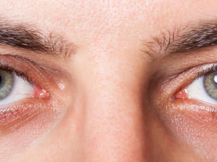
- Sudden-onset ocular pain, photophobia, tearing, blurred vision, ciliary flush.
- Anterior chamber cells/flare; fine KPs; posterior synechiae may develop rapidly.
- Typically unilateral and recurrent; IOP may be low initially, then variable.
- Granulomatous anterior uveitis (eg, sarcoidosis, TB)
- Blurred vision, mild discomfort; large “mutton-fat” KPs; iris nodules (Koeppe, Busacca).
- Often chronic, may be bilateral.
- Herpetic anterior uveitis (HSV/VZV)
- Unilateral; sectoral iris atrophy, elevated IOP disproportionate to cells, reduced corneal sensation; concurrent or prior keratitis, skin rash in VZV [1,3].
- JIA-associated uveitis
- Often asymptomatic; chronic, bilateral; band keratopathy, cataract, posterior synechiae.
- High amblyopia risk in young children [1].
- Traumatic iritis
- History of blunt trauma; photophobia and pain within 24–72 hours; cells/flare; cycloplegia relieves pain.
Complications
- Posterior synechiae, seclusion pupillae, iris bombe with secondary angle-closure glaucoma.
- Secondary open-angle glaucoma (trabeculitis, steroid response).
- Cataract (particularly posterior subcapsular).
- Cystoid macular edema; epiretinal membrane.
- Band keratopathy (chronic disease).
- Hypotony and phthisis bulbi (rare, severe chronic inflammation).
- Amblyopia in children.
Diagnosis
Clinical evaluation
- History: onset, laterality, systemic symptoms (back pain, psoriasis, oral/genital ulcers, diarrhea), infections, trauma, prior episodes, medications, travel/exposure.
- Examination:
- Visual acuity; IOP measurement.
- Slit-lamp: anterior chamber cells and flare (SUN grading), KPs, posterior synechiae, iris nodules; check for corneal involvement.
- Dilated fundus exam to assess vitreous cells and macular edema.
Imaging and laboratory testing
-
Test selection is phenotype- and history-driven; avoid indiscriminate panels in a first, mild, unilateral, nongranulomatous episode [1].
-
Common targeted tests:
- HLA-B27 typing for recurrent acute unilateral disease [1,2].
- Syphilis serology (nontreponemal plus treponemal tests) in most nontraumatic uveitis [1].
- Chest imaging and TB testing (IGRA) if granulomatous disease or risk factors [1].
- Lyme serology only with compatible exposure and systemic features [1].
- HSV/VZV suspicion: clinical diagnosis; consider aqueous PCR in atypical or refractory cases [1,3].
- OCT macula for cystoid macular edema; ultrasound B-scan if media opacity.
-
Coding: ICD-10-CM Iridocyclitis
- H20.0x Acute and subacute iridocyclitis (with laterality subcodes).
- H20.1x Chronic iridocyclitis (with laterality subcodes).
Differential diagnosis
| Entity | Distinguishing features |
|---|---|
| Bacterial/viral conjunctivitis | Diffuse conjunctival injection, discharge; no photophobia/true ciliary flush; no anterior chamber cells. See bacterial conjunctivitis. |
| Keratitis (infectious or inflammatory) | Corneal infiltrate/defect, focal staining, severe photophobia; may have anterior chamber reaction; pain with blinking. See keratitis. |
| Acute angle-closure glaucoma | Severe pain, headache, halos, nausea; mid-dilated fixed pupil; markedly elevated IOP; corneal edema; shallow chamber. |
| Episcleritis | Sectoral redness, mild tenderness, blanches with phenylephrine; no photophobia/cells. |
| Scleritis | Severe deep boring pain, violaceous hue, tenderness; may be associated with systemic autoimmune disease. |
| Endophthalmitis | Severe pain, hypopyon, decreased red reflex after surgery/penetration; vitritis prominent; emergent. |
| Dry eye disease | Burning, fluctuating blur, reflex tearing; no anterior chamber cells. See dry eye syndrome. |
| Hordeolum/chalazion | Localized lid nodule/tenderness; no intraocular inflammation. See chalazion. |
Treatment
Medical management
-
Goals: extinguish intraocular inflammation, relieve pain/photophobia, prevent synechiae and complications, treat underlying cause.
-
Topical corticosteroids (first-line unless contraindicated) [1,2]
- Prednisolone acetate 1%: 1 drop q1–2h while awake for severe, then taper to qid and slowly over weeks based on response.
- Difluprednate 0.05%: 1 drop qid for severe; taper cautiously; monitor IOP closely.
- Loteprednol 0.5%: 1 drop qid for milder disease or steroid responders.
-
Cycloplegic/mydriatic agents (pain relief; prevent synechiae) [1]
- Cyclopentolate 1%: 1 drop bid–tid.
- Homatropine 5%: 1 drop bid–tid.
- Atropine 1%: 1 drop qd–bid in severe fibrinous uveitis.
-
Intraocular pressure control [1,2]
- Topical beta-blocker (timolol 0.5% 1 drop bid), alpha-2 agonist (brimonidine 0.2% bid–tid), topical carbonic anhydrase inhibitor (dorzolamide 2% tid).
- Short-course oral acetazolamide 250 mg qid (or 500 mg ER bid) for marked IOP elevation, if no contraindications.
- Avoid miotics. Prostaglandin analogs are generally avoided in active uveitis due to potential to exacerbate inflammation; use cautiously if needed after quieting.
-
Infectious etiologies (add pathogen-directed therapy)
- Herpetic anterior uveitis (off-label dosing) [1,3]
- HSV: valacyclovir 500 mg PO tid or acyclovir 400 mg PO five times daily for 7–10 days; consider prophylaxis (eg, valacyclovir 500 mg PO bid) for recurrent disease. Use topical corticosteroid with concurrent antiviral.
- VZV: valacyclovir 1,000 mg PO tid for 7–10 days (initiate within 72 hours of rash when possible); longer courses often needed when uveitis is prominent; add topical corticosteroid and cycloplegic.
- Syphilis: treat per CDC neurosyphilis/ocular syphilis protocols in consultation with infectious diseases; topical steroids adjunctively once penicillin initiated.
- TB uveitis: coordinate antituberculous therapy with infectious diseases; topical/systemic steroids as indicated after initiating antimycobacterial treatment.
- CMV anterior uveitis: options include topical ganciclovir 0.15% gel and/or oral valganciclovir; management is off-label and specialist-guided [2].
-
Steroid-sparing/systemic therapy for chronic, recurrent, or bilateral noninfectious disease [1,2]
- Conventional immunomodulatory therapy (IMT): methotrexate, mycophenolate mofetil, azathioprine; dosing and monitoring per rheumatology/uveitis specialist.
- Biologics: adalimumab is FDA-approved for noninfectious intermediate, posterior, and panuveitis; may be used when anterior uveitis is part of a systemic uveitis phenotype or refractory to conventional IMT [6]. Off-label use for isolated anterior uveitis should be specialist-directed.
-
Monitoring
- Recheck 48–72 hours after initiation in moderate–severe cases; adjust frequency. Taper steroids slowly to avoid rebound. Monitor IOP for steroid response.
Procedural and surgical management
- Periocular corticosteroid injection (eg, posterior sub-Tenon triamcinolone acetonide 40 mg/mL, 0.5 mL) for refractory anterior segment inflammation or macular edema when infection excluded [1,2].
- Intravitreal corticosteroids (eg, triamcinolone 2–4 mg; dexamethasone implant 0.7 mg) mainly for posterior segment involvement/CME; selected refractory cases [1].
- Synechiolysis during dilation or surgically for extensive posterior synechiae impairing vision or pre-cataract surgery.
- Peripheral iridectomy for iris bombe with pupillary block once inflammation is controlled.
- Glaucoma surgery (eg, trabeculectomy, drainage devices) for uncontrolled IOP after inflammation is adequately suppressed.
- Cataract extraction when inflammation is quiescent (generally ≥3 months) with perioperative steroid optimization.
Special populations
- Pediatric (JIA-associated)
- Often asymptomatic; requires regular slit-lamp screening per pediatric rheumatology/ophthalmology guidelines; early IMT (eg, methotrexate) reduces complications [7].
- Aggressive control prevents amblyopia; coordinate with pediatric rheumatology.
- Pregnancy/lactation
- Avoid teratogenic IMT (eg, methotrexate, mycophenolate). Many topical agents and periocular steroids can be used with obstetric input; defer nonurgent systemic therapy changes to specialists.
- Immunocompromised
- Higher prevalence of infectious causes (eg, CMV); prioritize pathogen identification; avoid corticosteroid monotherapy.
- Older adults
- Higher risk of steroid-induced ocular hypertension and cataract; monitor IOP and lens status closely.
Prognosis
Acute nongranulomatous iridocyclitis typically responds to topical corticosteroids and cycloplegics within days, with resolution over 4–6 weeks. Recurrence is common, particularly in HLA-B27–associated disease. Chronic or granulomatous phenotypes and herpetic etiologies carry higher risks of cataract, glaucoma, and macular edema; long-term visual outcomes depend on timely control and complication management [1,2,5].
Prevention and Patient Counseling
- Adhere to prescribed drops and follow-up; do not stop steroids abruptly without guidance.
- Wear protective eyewear during activities with risk of ocular trauma to reduce traumatic iritis.
- Promptly treat systemic infections and coordinate care for autoimmune disease.
- Monitor IOP regularly during and after steroid therapy.
- Vaccination against herpes zoster (Shingrix) is recommended for adults ≥50 years and may reduce risk of herpes zoster ophthalmicus–related uveitis [8].
- Avoid contact lens wear during active inflammation.
When to Seek Immediate Care
- Sudden decrease in vision, severe eye pain, or intense light sensitivity.
- Seeing halos around lights with headache, nausea, or vomiting.
- Eye redness with new floaters, flashes, or a curtain over vision.
- A fixed mid-dilated pupil or markedly blurred vision.
- Eye injury or recent eye surgery with worsening pain or discharge.
References
- American Academy of Ophthalmology. Uveitis and Ocular Inflammation Preferred Practice Pattern. San Francisco, CA: AAO; 2019.
- EyeWiki. Anterior Uveitis. American Academy of Ophthalmology. https://eyewiki.org/Anterior_Uveitis. Accessed July 24, 2026.
- Miserocchi E, Fogliato G, Bianchi I, Bandello F, Modorati G. Clinical features of ocular herpetic infection in an Italian referral center. J Ophthalmol. 2014;2014:1-7. doi:10.1155/2014/906965.
- Jabs DA, Nussenblatt RB, Rosenbaum JT; Standardization of Uveitis Nomenclature (SUN) Working Group. Standardization of uveitis nomenclature for reporting clinical data. Am J Ophthalmol. 2005;140(3):509-516. doi:10.1016/j.ajo.2005.03.057.
- Gritz DC, Wong IG. Incidence and prevalence of uveitis in Northern California; the Northern California Epidemiology of Uveitis Study. Ophthalmology. 2004;111(3):491-500. doi:10.1016/j.ophtha.2003.06.014.
- U.S. Food and Drug Administration. HUMIRA (adalimumab) prescribing information. https://www.accessdata.fda.gov/drugsatfda_docs/label/2016/125057s399lbl.pdf. Accessed July 24, 2026.
- Angeles-Han ST, Ringold S, Beukelman T, et al. 2019 American College of Rheumatology/Arthritis Foundation guideline for the screening, monitoring, and treatment of juvenile idiopathic arthritis–associated uveitis. Arthritis Rheumatol. 2019;71(6):864-877. doi:10.1002/art.40885.
- Centers for Disease Control and Prevention. Shingles (Herpes Zoster) vaccination. https://www.cdc.gov/shingles/vaccination.html. Accessed July 24, 2026.
Disclaimer: This article is for informational purposes and is not a substitute for professional medical advice, diagnosis, or treatment. Always consult a qualified eye care professional about your specific condition. If you have sudden vision loss, severe eye pain, or an eye injury, seek emergency care immediately.