Uveitis is inflammation of the uveal tract (iris, ciliary body, choroid) and adjacent structures. It most often presents with eye pain, redness, light sensitivity, floaters, and/or decreased vision. Diagnosis is clinical by slit-lamp and dilated fundus examination, supported by targeted imaging and laboratory tests. Treatment depends on anatomic location and cause and includes corticosteroids, cycloplegics, immunomodulatory therapy, and antimicrobials for infectious etiologies; prompt control of inflammation prevents vision-threatening complications.
Key Points
- Uveitis is classified by anatomic site: anterior, intermediate, posterior, and panuveitis per Standardization of Uveitis Nomenclature (SUN) criteria [1].
- Infectious and noninfectious causes must be distinguished early; corticosteroids without antimicrobial coverage can worsen unrecognized infection (eg, syphilis, tuberculosis, herpetic disease) [2][3][4].
- First-line therapy for noninfectious anterior uveitis is topical corticosteroids plus a cycloplegic; posterior-segment disease often requires periocular/intravitreal steroids, systemic corticosteroids, and steroid-sparing immunomodulators [2].
- FDA-approved agents for noninfectious posterior-segment uveitis include intravitreal dexamethasone and fluocinolone implants and systemic adalimumab [5][6][7].
- Cystoid macular edema, cataract, and glaucoma are the leading causes of visual morbidity; vigilant monitoring with OCT and intraocular pressure (IOP) checks is essential [2].
- Laboratory testing is selective and led by history and exam; “shotgun” panels are low yield and can mislead [2].
Anatomy and Physiology
The uvea comprises:
- Iris: smooth muscle diaphragm regulating light entry via the pupil.
- Ciliary body: produces aqueous humor and controls accommodation; inflammation impairs aqueous dynamics and can lower or raise IOP.
- Choroid: vascular layer supplying outer retina; inflammation affects photoreceptors and retinal pigment epithelium.
Adjacent structures commonly involved include the corneal endothelium (keratic precipitates), lens (posterior subcapsular cataract), vitreous (vitritis), retina (retinitis/vasculitis), and optic nerve (papillitis).
Etiology
- Noninfectious/immune-mediated
- HLA-B27–associated spondyloarthropathies (ankylosing spondylitis, reactive arthritis, psoriatic arthritis, IBD-related)
- Sarcoidosis
- Behçet disease
- Vogt–Koyanagi–Harada (VKH) disease
- Birdshot chorioretinopathy
- Juvenile idiopathic arthritis (JIA)
- Idiopathic
- Infectious
- Herpes simplex virus (HSV) and varicella-zoster virus (VZV)
- Cytomegalovirus (CMV; retinitis in immunocompromised; anterior uveitis in some immunocompetent)
- Toxoplasma gondii (necrotizing retinochoroiditis)
- Treponema pallidum (syphilis)
- Mycobacterium tuberculosis (TB)
- Borrelia burgdorferi (Lyme disease; regional)
- Bartonella henselae (neuroretinitis)
- Endogenous or postoperative endophthalmitis (bacterial/fungal)
- Masquerade syndromes
- Primary intraocular lymphoma, leukemia
- Retinal degenerations, intraocular foreign body
- Lens-induced and trauma/surgery-related
- Phacoantigenic (phacogenic) uveitis, phacolytic glaucoma
- Sympathetic ophthalmia
- Drug-induced
- Cidofovir, rifabutin, bisphosphonates, sulfonamides, immune checkpoint inhibitors [2]
Pathophysiology
Innate and adaptive immune responses target uveal and retinal antigens or pathogen components, disrupting the blood–aqueous and blood–retinal barriers. Cytokine cascades (eg, TNF-α, IL-6) drive leukocyte recruitment into the anterior chamber and vitreous. Chronic inflammation promotes fibrocellular proliferation, synechiae, trabecular meshwork damage (secondary glaucoma), lens epithelial changes (posterior subcapsular cataract), and macular microvascular leakage (cystoid macular edema).
Epidemiology
- Uveitis predominantly affects working-age adults but occurs in children (notably JIA-associated) and older adults (eg, herpetic, sarcoid) [2][8].
- US incidence and prevalence estimates vary by study; uveitis is a leading cause of preventable visual loss and legal blindness in the United States [8].
Classification
Per SUN Working Group [1]:
- By anatomic location
- Anterior: primary site in anterior chamber (iritis, iridocyclitis, anterior cyclitis)
- Intermediate: vitreous/pars plana (pars planitis)
- Posterior: retina/choroid (retinitis, choroiditis, retinochoroiditis)
- Panuveitis: anterior chamber, vitreous, and posterior segment all inflamed
- By onset: sudden vs insidious
- By duration: limited (< 3 months) vs persistent (≥ 3 months)
- By course: acute, recurrent, or chronic
- By laterality: unilateral, alternating, bilateral
Symptoms and Signs
- Anterior uveitis (most common)
- Symptoms: ocular pain, redness, photophobia, blurred vision.
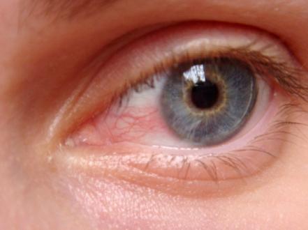
- Signs: ciliary flush, anterior chamber cells/flare, keratic precipitates (non-granulomatous vs granulomatous “mutton-fat”), posterior synechiae (irregular pupil), hypopyon (eg, HLA-B27), variable IOP (low in acute inflammation; elevated in herpetic or steroid response).
- Intermediate uveitis
- Symptoms: floaters, blurred vision; minimal pain/redness.
- Signs: vitritis (cells), “snowballs” and “snowbanking” at pars plana, peripheral vascular sheathing; CME common.
- Posterior uveitis
- Symptoms: painless vision loss, scotomas, floaters, photopsias.
- Signs: retinitis/choroiditis lesions, retinal vasculitis, vitritis, optic disc edema, serous retinal detachment; modality-specific angiographic leakage.
- Panuveitis
- Combination of anterior and posterior findings; consider VKH, sarcoidosis, sympathetic ophthalmia, Behçet disease.
Complications
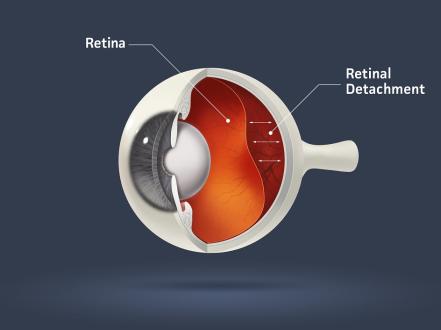
- Ocular: cystoid macular edema, epiretinal membrane, posterior subcapsular cataract, glaucoma/ocular hypertension, band keratopathy, posterior and peripheral anterior synechiae, hypotony, choroidal neovascularization, retinal detachment, optic neuropathy.
- Treatment-related: steroid-induced ocular hypertension/glaucoma and cataract; periocular/intravitreal injection risks (infection, hemorrhage, detachment); systemic immunosuppression toxicities (hepatic, hematologic, renal, infectious).
Diagnosis
Clinical evaluation
- History: symptom chronology, laterality, prior episodes, systemic symptoms (oral/genital ulcers, back pain, rash, cough/dyspnea, neurologic/auditory changes, GI symptoms, tick exposure, TB exposure), medications, surgeries/trauma, sexual history and risk.
- Examination:
- Visual acuity, pupils (consensual photophobia, synechiae), IOP.
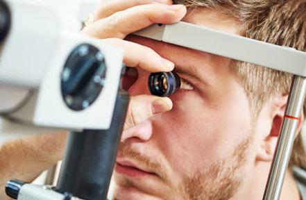
- Slit-lamp: corneal endothelium (KPs), anterior chamber cell and flare grading, hypopyon/hyphema, posterior synechiae, lens changes.
- Dilated fundus exam: vitreous cells/haze, retinal/choroidal lesions, vasculitis, macular status, optic nerve head.
- Avoid corticosteroids until major infections (syphilis, TB, herpetic disease) are reasonably excluded or covered [2][3][4].
Imaging and laboratory testing
- Imaging
- Optical coherence tomography (OCT): macular edema, epiretinal membrane.
- Fluorescein angiography: macular leakage, vasculitis, optic disc leakage.
- Indocyanine green angiography: choroidal inflammatory lesions (eg, birdshot).
- B-scan ultrasonography: when media are opaque.
- Chest radiograph/CT: sarcoidosis, TB (as indicated).
- Laboratory testing (targeted)
- Syphilis serology (treponemal + non-treponemal) in most new uveitis cases [3].
- Interferon-γ release assay (IGRA) for TB if risk factors/exam suggestive [4].
- Serum ACE/lysozyme and chest imaging for suspected sarcoid; interpret cautiously due to limited specificity [2].
- HLA-B27 typing for recurrent acute anterior uveitis.
- Toxoplasma IgG/IgM with compatible necrotizing retinitis.
- ANA and related testing in pediatric cases (JIA risk stratification).
- HIV testing when posterior uveitis/CMV retinitis is suspected or systemic risk exists.
- Aqueous/vitreous PCR when infectious posterior uveitis is uncertain (HSV/VZV/CMV, T. gondii).
Differential diagnosis
| Entity | Distinguishing features |
|---|---|
| Bacterial conjunctivitis | Diffuse conjunctival injection, purulent discharge, minimal photophobia, normal pupil/IOP; no anterior chamber cells |
| Keratitis (infectious) | Corneal epithelial defect/infiltrate, severe photophobia, pain; positive corneal staining; risk with contact lens wear |
| Scleritis | Severe, boring pain radiating to face; globe tenderness; deep episcleral vessel involvement; often systemic autoimmune disease |
| Acute angle-closure glaucoma | Sudden severe pain, halos, headache/nausea; mid-dilated fixed pupil; markedly elevated IOP; corneal edema |
| Endophthalmitis | Postoperative or endogenous; severe pain, profound vision loss, hypopyon, dense vitritis; emergent |
| Chalazion | Focal eyelid nodule; no photophobia, no intraocular inflammation |
| Dry eye syndrome | Burning/grittiness, fluctuating vision, worse with reading/screens; minimal photophobia; no intraocular cells |
Treatment
Therapy depends on anatomic location and cause. Rule out/treat infection before or alongside corticosteroids and immunosuppression.
Medical management
- Noninfectious anterior uveitis
- Topical corticosteroids: prednisolone acetate 1% q1–2h while awake, then taper to qid and slower based on cell grade; or difluprednate 0.05% qid then taper [2].
- Cycloplegics/mydriatics: homatropine 5% bid–tid, cyclopentolate 1% tid, or atropine 1% qd–bid to relieve ciliary spasm and prevent synechiae.
- IOP control (if elevated): topical β-blocker (timolol 0.5% bid), α-agonist (brimonidine 0.2% tid), or carbonic anhydrase inhibitor (dorzolamide 2% tid); prostaglandin analogs are generally avoided during active uveitis.
- Periocular corticosteroid (sub-Tenon triamcinolone acetonide 40 mg) for refractory unilateral disease.
- Intermediate, posterior, and panuveitis (noninfectious)
- Systemic corticosteroids: prednisone 0.5–1 mg/kg/day with taper; for sight-threatening inflammation, IV methylprednisolone 1 g/day for 1–3 days [2].
- Local steroid therapy: periocular triamcinolone; intravitreal triamcinolone (2–4 mg); intravitreal dexamethasone implant 0.7 mg (Ozurdex, FDA-approved for noninfectious uveitis affecting the posterior segment) [5]; fluocinolone acetonide implants 0.18 mg (Yutiq) or 0.59 mg (Retisert) for chronic noninfectious posterior uveitis [6].
- Steroid-sparing immunomodulatory therapy (off-label unless noted): methotrexate 10–25 mg weekly with folic acid; mycophenolate mofetil 1,000–1,500 mg bid; azathioprine 1–2 mg/kg/day; cyclosporine 2–5 mg/kg/day; others per subspecialist [2].
- Biologic therapy: adalimumab (Humira) 80 mg loading, then 40 mg SC every 2 weeks; FDA-approved for noninfectious intermediate, posterior, and panuveitis based on VISUAL trials [7][9]. Infliximab is used off-label in select entities (eg, Behçet).
- Infectious uveitis (examples; treat with or in consultation with infectious disease/uveitis specialist)
- Herpetic anterior uveitis: valacyclovir 1 g PO tid or acyclovir 800 mg PO five times daily for 7–10 days, then maintenance as indicated; add topical steroids once antiviral therapy is started [2].
- Ocular toxoplasmosis: pyrimethamine (loading 50–75 mg/day, then 25–50 mg/day) plus sulfadiazine 1–1.5 g qid with leucovorin 10–25 mg 3x/week; add prednisone (eg, 20–60 mg/day) 24–48 hours after antibiotics start; TMP–SMX 160/800 mg bid is a common alternative [2].
- Syphilitic uveitis: treat as neurosyphilis with aqueous crystalline penicillin G 18–24 million units/day IV for 10–14 days per CDC guidelines [3].
- TB-associated uveitis: multi-drug anti-TB therapy per CDC/ATS/IDSA guidelines; add corticosteroids under cover as indicated [4].
- CMV retinitis (immunocompromised): valganciclovir 900 mg PO bid for 14–21 days, then 900 mg daily; intravitreal ganciclovir or foscarnet adjuncts; optimize antiretroviral therapy when applicable [2].
Procedural and surgical management
- Periocular/intravitreal steroid injections or implants for localized refractory inflammation or CME (see above).
- Laser/surgical interventions for complications:
- Peripheral iridectomy for iris bombe from posterior synechiae; synechiolysis during cataract surgery as needed.
- Glaucoma procedures (trabeculectomy, tubes) for uncontrolled uveitic glaucoma.
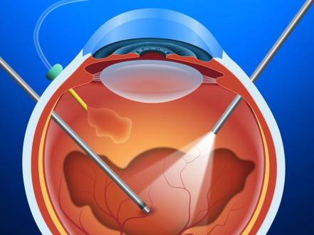
- Pars plana vitrectomy for nonresolving vitreous opacities, traction, diagnostic biopsy (lymphoma), or infectious source control.
- Cataract extraction after sustained quiescence (commonly ≥ 3 months) with perioperative inflammation control.
Special populations
- Pediatric (eg, JIA-associated)
- Often asymptomatic; requires routine screening and aggressive control to prevent amblyopia and complications [2].
- Use the lowest effective topical steroid burden; early steroid-sparing IMT is common.
- Pregnancy/lactation
- Favor local therapies and systemic corticosteroids at the lowest effective dose.
- Avoid teratogens (methotrexate, mycophenolate mofetil); azathioprine may be considered with obstetric input; adalimumab has pregnancy registry data but timing near delivery requires planning; most pass into breast milk variably—specialist oversight required.
- Immunocompromised/elderly
- Higher likelihood of infectious uveitis (eg, CMV retinitis); broaden diagnostic workup.
- Monitor closely for medication toxicities and steroid-induced IOP elevation and cataract.
Prognosis
Outcome depends on etiology, anatomic location, promptness of control, and treatment adherence. Many cases of anterior uveitis respond rapidly to topical therapy but may recur, especially in HLA-B27 disease. Intermediate/posterior/panuveitis often require prolonged therapy; steroid-sparing immunomodulators and adalimumab reduce relapses and treatment failures in noninfectious posterior-segment uveitis [7][9]. Macular edema, glaucoma, and cataract drive long-term visual outcomes.
Prevention and Patient Counseling
- Adhere to the prescribed regimen and taper schedule; abrupt steroid cessation risks rebound.
- Report new floaters, photopsias, or vision changes promptly; keep scheduled OCT/IOP checks.
- Avoid contact lenses during active inflammation.
- Manage systemic diseases (eg, spondyloarthropathy, sarcoidosis) in coordination with rheumatology.
- Vaccination with recombinant zoster vaccine (Shingrix) reduces shingles and may lower risk of VZV-associated ocular inflammation; timing should be coordinated with immunosuppression.
When to Seek Immediate Care
- Sudden vision loss, a dark curtain over vision, or many new floaters/flashes
- Severe eye pain with redness or headache, nausea, and halos around lights
- Pupil that will not move or a markedly different eye pressure sensation
- Symptoms after eye surgery or penetrating eye injury
- Eye symptoms with fever, rash, stiff neck, or new neurologic problems
- Any vision change in people with HIV/AIDS, on chemotherapy, or taking strong immunosuppressants
References
- Jabs DA, Nussenblatt RB, Rosenbaum JT; Standardization of Uveitis Nomenclature (SUN) Working Group. Standardization of uveitis nomenclature for reporting clinical data—2021 revision. Ocul Immunol Inflamm. 2021;29(1):1-16. doi:10.1080/09273948.2020.1798898.
- American Academy of Ophthalmology. Preferred Practice Pattern: Uveitis—Initial Evaluation and Management. San Francisco, CA: AAO; 2019/2020.
- Centers for Disease Control and Prevention (CDC). Sexually Transmitted Infections Treatment Guidelines, 2021—Ocular Syphilis. https://www.cdc.gov/std/treatment-guidelines/syphilis.htm
- Nahid P, Dorman SE, Alipanah N, et al. Official ATS/CDC/IDSA Clinical Practice Guidelines: Treatment of Drug-Susceptible Tuberculosis. Clin Infect Dis. 2016;63(7):e147-e195. doi:10.1093/cid/ciw376.
- FDA/DailyMed. Ozurdex (dexamethasone intravitreal implant) label. https://dailymed.nlm.nih.gov/dailymed/drugInfo.cfm?setid=8a1a3a8e-9d2c-4a3a-8e1a-8a9b8a3a8e9d
- FDA/DailyMed. Yutiq (fluocinolone acetonide intravitreal implant) label. https://dailymed.nlm.nih.gov/dailymed/drugInfo.cfm?setid=77e2aa32-69c1-4e26-8c7f-7f1a2b4a1b1c
- Humira (adalimumab) Prescribing Information—Noninfectious Uveitis Indication. https://www.rxabbvie.com/pdf/humira.pdf
- National Eye Institute (NEI). Facts About Uveitis. https://www.nei.nih.gov/learn-about-eye-health/eye-conditions-and-diseases/uveitis
- Nguyen QD, Merrill PT, Jaffe GJ, et al. Adalimumab for prevention of uveitic flare in patients with inactive non-infectious uveitis (VISUAL II). Lancet. 2016;388(10050):1183-1192. doi:10.1016/S0140-6736(16)31339-3.
This article is for informational purposes and is not a substitute for professional medical advice, diagnosis, or treatment. Always consult a qualified eye care professional about your specific condition. If you have sudden vision loss, severe eye pain, or an eye injury, seek emergency care immediately.