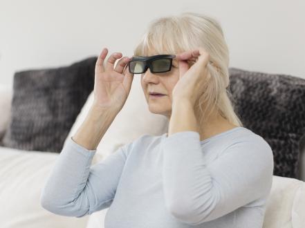
Night blindness (nyctalopia) is impaired vision under low-light or dark conditions due to reduced rod photoreceptor function, media opacity, or optical factors. Patients report difficulty seeing at dusk or in dim rooms, slow dark adaptation, and problems driving at night. Diagnosis is clinical and supported by dark adaptometry, electroretinography, retinal imaging, and, when indicated, serum vitamin A testing. Treatment targets the cause (eg, vitamin A repletion, cataract surgery, discontinuation of retinotoxic drugs, genetic counseling and low-vision support for inherited retinal dystrophies).
Key Points
- Night blindness is a symptom, not a diagnosis; common etiologies include vitamin A deficiency, inherited rod-dominant retinal disorders (eg, retinitis pigmentosa), medication toxicity (eg, isotretinoin), and light scatter from cataract.
- Photopic visual acuity may be normal; targeted history (dark adaptation, driving at night) and scotopic testing reveal deficits.
- Vitamin A deficiency is uncommon in the US but should be considered in malnutrition or fat malabsorption; timely repletion can reverse symptoms.
- Electroretinography (ERG) shows reduced scotopic responses in rod dysfunction; fundus exam and multimodal imaging help distinguish dystrophies (eg, bone spicule pigment in retinitis pigmentosa).
- Avoid empiric high-dose vitamin A in inherited retinal degenerations without specialist guidance due to safety concerns and uncertain benefit.
- Counsel on night driving safety; address reversible contributors (glare control, cataract, refractive error, ocular surface disease).
Anatomy and Physiology
Rods mediate scotopic (low-light) vision and peripheral motion detection. Rod density peaks in the mid-peripheral retina; the fovea is cone-dominant. Rhodopsin (rod visual pigment) requires retinaldehyde derived from vitamin A (retinol). The retinal pigment epithelium (RPE) supports photoreceptor function by recycling chromophore and phagocytosing outer segments. Disruption of rods, the visual cycle, or optical media transmission impairs dark adaptation and scotopic sensitivity.
Etiology
- Retinal/visual cycle
- Inherited retinal degenerations: retinitis pigmentosa (RP), rod-cone dystrophy, choroideremia, congenital stationary night blindness (CSNB), Oguchi disease.
- Acquired: vitamin A deficiency (malnutrition; fat malabsorption—celiac disease, Crohn disease, pancreatic insufficiency, cholestatic liver disease; post–bariatric surgery), retinal toxicity (eg, isotretinoin), retinal iron deposition (siderosis bulbi).
- Media and optical factors
- Cataract (nuclear sclerosis, posterior subcapsular) increasing light scatter and reducing contrast.
- Uncorrected refractive error (including night myopia), irregular astigmatism (eg, keratoconus), and higher-order aberrations (eg, post-refractive surgery).
- Ocular surface disease (dry eye) causing tear-film instability and glare.
- Neurologic/other
- Advanced glaucoma and optic neuropathies can reduce contrast sensitivity; true nyctalopia is typically retinal.
Medications associated with nyctalopia or impaired dark adaptation: isotretinoin, phenothiazines, chloroquine/hydroxychloroquine (retinal toxicity), sildenafil-class phosphodiesterase inhibitors [1][2]. Mechanisms and causality vary; medication review is essential.
Pathophysiology
- Rod dysfunction: Loss or dysfunction of rods reduces scotopic ERG amplitude and elevates scotopic thresholds, prolonging dark adaptation (eg, RP).
- Visual cycle impairment: Inadequate retinoid availability (vitamin A deficiency; RPE65 or other cycle gene defects) limits rhodopsin regeneration, causing delayed dark adaptation.
- Light scatter and aberrations: Cataract and irregular optics degrade retinal image quality, disproportionately affecting low-luminance and mesopic tasks by reducing contrast and increasing glare.
- Secondary effects: Chronic ocular surface disease destabilizes the tear film, increasing straylight and reducing low-luminance performance.
Epidemiology
- Retinitis pigmentosa affects approximately 1 in 4,000 in the US; nyctalopia is often the first symptom [3].
- Vitamin A deficiency is rare in the US but occurs in patients with malabsorption, liver disease, or restrictive diets; prevalence data are limited in high-income settings.
- Cataract prevalence rises with age; night driving complaints are common and often improve after surgery [4].
- Procedure-related complaints (night glare/halos) can follow corneal refractive surgery; most attenuate over months.
Classification
- By cause
- Retinal/visual cycle (rod dysfunction): inherited (RP, CSNB, Oguchi, choroideremia); acquired (vitamin A deficiency, toxicity).
- Optical/media: cataract, refractive error/night myopia, aberrations (keratoconus, post-surgery), ocular surface disease.
- By onset
- Congenital/early-onset (CSNB, Oguchi).
- Acquired (deficiency, cataract, toxicity, degenerations).
- By reversibility
- Potentially reversible (deficiency, cataract, refractive/optical, medication-induced).
- Typically progressive (RP, choroideremia).
Symptoms and Signs
- General
- Difficulty seeing in dim light; slow dark adaptation after bright exposure.
- Problems driving at dusk/night (poor peripheral detection, delayed recovery from oncoming headlights).
- Normal or near-normal photopic visual acuity early; constricted peripheral fields in rod-cone dystrophies.
- Variant-specific
- Vitamin A deficiency: xerosis (dry conjunctiva/cornea), Bitot spots, decreased scotopic sensitivity, reduced color saturation; severe cases show corneal ulcers/keratomalacia.
- Retinitis pigmentosa: nyctalopia, peripheral field loss, fundus with bone spicule pigmentation, attenuated vessels, waxy disc pallor.
- CSNB/Oguchi: lifelong night blindness, normal central acuity; Oguchi shows Mizuo–Nakamura fundus sheen that normalizes after dark adaptation.
- Cataract: glare, halos, reduced contrast; worse under backlit/night conditions.
- Optical/ocular surface (keratoconus, dry eye, post-refractive): ghosting, starbursts, fluctuating blur, glare.
Complications
- Reduced driving safety and mobility at night; increased fall risk in low illumination.
- Progressive peripheral visual field constriction in rod-cone dystrophies leading to legal blindness.
- Corneal complications in severe vitamin A deficiency (ulceration, scarring).
- Psychosocial impact: activity limitation, anxiety about night travel.
Diagnosis
Clinical evaluation
- History: onset and time course; dark adaptation difficulty; night driving performance; glare sensitivity; peripheral field symptoms; diet; gastrointestinal or hepatic disease (malabsorption); bariatric surgery; medications (isotretinoin, chloroquine derivatives, phenothiazines, PDE-5 inhibitors); family history of retinal disease; consanguinity.
- Examination:
- Visual acuity (photopic) and pinhole, refraction (assess night myopia/uncorrected error).
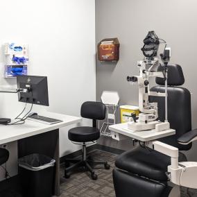
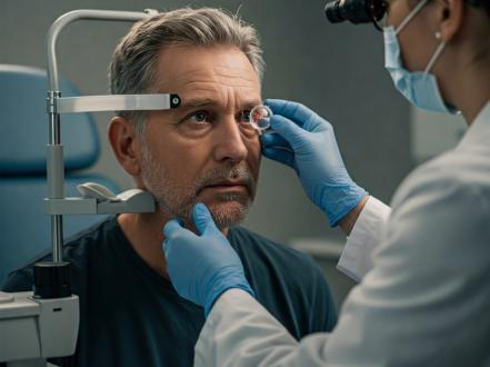
- Slit-lamp for ocular surface disease and cataract grade.
- Dilated fundus examination for pigmentary changes, vessel attenuation, RPE alterations.
- Confrontation fields; formal perimetry when indicated.
Imaging and laboratory testing
- Dark adaptometry: rod-mediated dark adaptation curve; prolonged rod intercept time in visual cycle/rod disorders [5].
- Electroretinography (ERG): reduced scotopic a- and b-waves in rod dysfunction; photopic responses relatively preserved early in rod-cone disease [6].
- Optical coherence tomography (OCT): outer retinal/ellipsoid zone loss in degenerations; macular complications (eg, cystoid macular edema in RP).
- Fundus autofluorescence: rings/patches indicating RPE/photoreceptor stress or loss.
- Visual fields (Goldmann or automated): mid-peripheral constriction in RP; ring scotomas.
- Laboratory testing:
- Serum retinol (vitamin A) for suspected deficiency; assess fat-soluble vitamin status and underlying malabsorption.
- Genetic testing for inherited retinal degenerations per AAO PPP recommendations [6].
- Ancillary:
- Consider medication levels or collaboration with prescribers for suspected toxicity.
- ERG and dark adaptometry before/after medication changes when feasible.
CPT examples: dark adaptation (92284), full-field ERG (92273), multifocal ERG (92274), OCT retina (92134), fundus photography (92250), perimetry (92083).
Differential diagnosis
| Entity | Distinguishing features |
|---|---|
| Vitamin A deficiency | History of malabsorption, restrictive diet, xerosis/Bitot spots; low serum retinol; rapid improvement with repletion |
| Retinitis pigmentosa | Family history; bone spicule pigment, attenuated arterioles; scotopic ERG reduced; progressive field constriction |
| Congenital stationary night blindness | Lifelong symptoms; normal fundus (except Oguchi sheen); stable acuity; characteristic ERG patterns |
| Choroideremia | X-linked; male patients; choroidal/RPE atrophy beginning peripherally; progressive field loss |
| Cataract (PSC/nuclear) | Glare/halos; lens opacity on slit-lamp; improvement after cataract surgery |
| Night myopia/uncorrected refractive error | Worse low-light distance vision improved with updated refraction; normal retina |
| Keratoconus/irregular astigmatism | Scissoring reflex; corneal topography abnormal; ghosting/halos; variable acuity |
| Post-refractive surgery aberrations | Night glare/starbursts; higher-order aberrations on aberrometry; often improve over time |
| Ocular surface disease (dry eye) | Fluctuating blur, burning; tear-film instability; worsens with prolonged staring/air exposure |
| Medication toxicity (isotretinoin, etc.) | Temporal association with drug; ERG/dark adaptation changes; may improve after discontinuation |
| Optic neuropathy/glaucoma | Reduced contrast sensitivity; visual field defects not purely rod-mediated; normal scotopic ERG |
For ocular surface and infectious mimics causing glare, see dry eye syndrome (/blog/dry-eye-syndrome), keratitis (/disease/keratitis), and bacterial conjunctivitis (/disease/bacterial-conjunctivitis). Eyelid lesions (eg, chalazion, /disease/chalazion) can induce astigmatism and night glare.
Treatment
Medical management
- Vitamin A deficiency
- Adults: retinol (vitamin A) 25,000 to 50,000 IU PO daily for 1–2 weeks, then dietary maintenance and treatment of underlying malabsorption; severe xerophthalmia may require higher-dose, short-course regimens as per Merck Manual/WHO protocols [7][8].
- Children with xerophthalmia (Merck/WHO): 200,000 IU PO once daily for 2 days and again after 2 weeks (>12 months); 100,000 IU if 6–11 months; 50,000 IU if <6 months [7][8].
- Pregnancy/lactation: avoid high-dose vitamin A; consult obstetrics; use lower, carefully supervised dosing [7][8].
- Monitor liver function in prolonged/high-dose therapy; avoid chronic megadoses due to hepatotoxicity and teratogenicity.
- Medication-induced nyctalopia
- Coordinate with the prescribing clinician to reduce dose or discontinue the offending agent (eg, isotretinoin); document baseline and follow-up dark adaptation/ERG when feasible [1].
- Retinal degenerations (RP, choroideremia, CSNB)
- No established pharmacologic reversal of nyctalopia; manage complications (eg, cystoid macular edema with topical or systemic carbonic anhydrase inhibitors) [6].
- Genetic counseling, low-vision rehabilitation, orientation and mobility training, and driving counseling.
- Gene therapy is FDA-approved for RPE65-associated disease (voretigene neparvovec) but targets severe early-onset forms, not typical adult-onset RP [6].
- Empiric high-dose vitamin A palmitate historically studied in RP; routine use is not recommended without specialist oversight due to safety concerns and mixed evidence [6].
- Optical/media causes
- Update refraction; consider night-driving glasses with anti-reflective coatings.
- Manage ocular surface disease (lubricants, lid hygiene, anti-inflammatory therapy as indicated).
- Address keratoconus with rigid gas-permeable or scleral lenses; consider corneal cross-linking in progressive disease (to stabilize, not directly treat nyctalopia).
Representative agents and doses (examples; individualized by clinician):
- Retinol (vitamin A) PO: deficiency, adults 25,000–50,000 IU daily × 1–2 weeks [7].
- Acetazolamide PO (for RP-associated cystoid macular edema): 250 mg bid–tid, off-label; monitor electrolytes/renal function [6].
All pharmacotherapy should follow FDA labeling when available; off-label uses are noted.
Procedural and surgical management
- Cataract surgery: indicated when lens opacity limits function; reliably improves glare and low-luminance performance in lens-related complaints [4].
- Corneal procedures: corneal cross-linking for progressive keratoconus (stabilization); topography-guided PRK or intracorneal ring segments in selected cases.
- Low-vision devices: telescopes, contrast-enhancing filters, task lighting; orientation/mobility training.
Special populations
- Pediatric
- Consider CSNB, Oguchi disease, early-onset RP; evaluate for nystagmus, myopia, and strabismus.
- Dose vitamin A per age/weight if deficiency; avoid adult megadoses [7][8].
- Genetic testing and counseling for inherited etiologies.
- Pregnancy/lactation
- Avoid high-dose vitamin A (teratogenic); manage deficiency with supervised, lower dosing and dietary measures [7][8].
- Night-driving counseling and environmental adaptations.
- Immunocompromised/liver disease/malabsorption
- Higher risk of deficiency; coordinate with gastroenterology/hepatology; parenteral vitamin A may be needed in severe malabsorption [7].
- Older adults
- Higher prevalence of cataract and ocular surface disease; polypharmacy review for drugs affecting dark adaptation.
Prognosis
- Vitamin A deficiency: often reversible when treated promptly; prolonged deficiency risks permanent retinal and corneal damage [7].
- Retinitis pigmentosa and related dystrophies: typically progressive; nyctalopia precedes peripheral field constriction and eventual central involvement; rate varies by genotype [3][6].
- Optical/media etiologies: favorable outcomes after addressing refractive error, ocular surface disease, or cataract.
- Medication-induced cases: partial or full recovery may occur after discontinuation; not guaranteed.
Prevention and Patient Counseling
- Maintain a balanced diet with adequate vitamin A (eg, dairy, eggs, liver, fortified foods; provitamin A carotenoids in leafy greens and orange vegetables).
- Seek evaluation for persistent night driving difficulty, especially with a family history of retinal disease or malabsorption history.
- Avoid high-dose vitamin A supplements without medical supervision.
- Optimize environmental lighting, use contrast-enhancing task lighting, and consider anti-reflective lenses.
- Follow state regulations and clinician advice about night driving; consider ride alternatives if unsafe.
When to Seek Immediate Care
- Sudden vision loss or a new dark curtain in vision.
- Severe eye pain, redness, or light sensitivity.
- New flashes of light or a sudden shower of floaters.
- Chemical injury or eye trauma.
- Rapid onset of night blindness after starting a new medication. Call 911 for severe eye injury or sudden, severe vision loss.
References
- Fraunfelder FW, Fraunfelder FT. Isotretinoin-associated night vision disturbances. J Am Acad Dermatol. 2004;50(3):476-480. doi:10.1016/j.jaad.2003.08.036.
- Marmor MF, Kellner U, Lai TYY, Lyons JS, Mieler WF. Recommendations on Screening for Chloroquine and Hydroxychloroquine Retinopathy (2016 Revision). Ophthalmology. 2016;123(6):1386-1394. doi:10.1016/j.ophtha.2016.01.058.
- Hartong DT, Berson EL, Dryja TP. Retinitis pigmentosa. Lancet. 2006;368(9549):1795-1809. doi:10.1016/S0140-6736(06)69740-7.
- Ehrlich JR, Oh E, Shim M, et al. Association of Cataract and Cataract Surgery With Nighttime Driving Difficulty Among Older Adults. JAMA Ophthalmol. 2019;137(9):967-973. doi:10.1001/jamaophthalmol.2019.2335.
- Jackson GR, Edwards JG. A short-duration dark adaptation test for age-related maculopathy. J Ocul Biol Dis Infor. 2008;1(1):7-11. doi:10.1007/s12177-008-9002-6.
- American Academy of Ophthalmology. Inherited Retinal Degenerations Preferred Practice Pattern. 2022. https://www.aao.org/preferred-practice-pattern/inherited-retinal-degenerations-ppp
- Merck Manual Professional Edition. Vitamin A Deficiency. Updated 2023. https://www.merckmanuals.com/professional/nutritional-disorders/vitamin-deficiency-dependency-and-toxicity/vitamin-a-deficiency
- World Health Organization. Vitamin A supplementation for xerophthalmia. Guideline. 2011. https://www.who.int/publications/i/item/9789241501767


This article is for informational purposes and is not a substitute for professional medical advice, diagnosis, or treatment. Always consult a qualified eye care professional about your specific condition. If you have sudden vision loss, severe eye pain, or an eye injury, seek emergency care immediately.