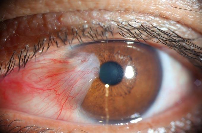
Pterygium (pterygos in Greek means wing) is a benign triangular fibrous growth growing from the inner corner of the eye to the cornea. Typically, pterygium grows on the nasal side, sometimes on the temporal side, but bilateral growth can also occur.
The exact cause of pterygium is unknown. However, the most studied risk factor is ultraviolet radiation. Ultraviolet light generates free radicals that damage the DNA, RNA, and extracellular matrix of cells. Ultraviolet light induces the expression of cytokines and growth factors in pterygium epithelial cells. Pterygium tissue also showed elevated levels of T cells and inflammatory markers compared to normal conjunctival tissue.
Pterygium occurs most often in sunny and windy climates. That’s why this disease is sometimes called "surfer's eye." The tendency to progress and rapid growth in most cases is seen in young people.
Risk factors of Pterygium
- Exposure to ultraviolet rays (the single most significant risk factor).
- Exposure to irritants (dust, sand, allergens, wind).
- Dry eye disease.
- Inflammatory processes.
Pterygium symptoms
Pterygium is a wedge-shaped translucent membrane with an apex extending to the cornea. It has a white or pinkish color, depending on the number of vessels. Pterygium can be asymptomatic without causing any complaints. The main symptoms are:
- redness of the eyes;
- irritation, foreign body feeling;
- tearing;
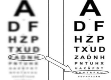
- decreased visual acuity with ingrowth into the optical zone of the cornea (regular or irregular astigmatism);
- degenerative cystic changes.
Pterygium Diagnosis
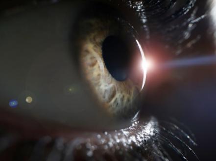
Diagnosis is made clinically based on slit-lamp examination and the typical appearance of the pathological process.
How is it treated?
Small pterygiums without visual impairment do not require treatment. If the eye is severely irritated or red, the ophthalmologist may prescribe artificial tears or eye ointments containing corticosteroids to reduce inflammation. But it is important to understand that medications do not affect the rate of progression of pterygium. It is recommended to visit an ophthalmologist every 6-12 months to monitor the process.
Pterygium surgical treatment
Indications for surgical removal of the pterygium:
- Decreased visual acuity due to astigmatism or growth in the center of the cornea.
- Cosmetically significant pterygium.
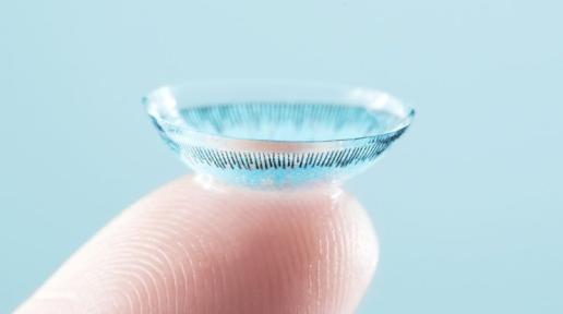
- When pterygium interferes with contact lens wear.
- Symptomatic degenerative changes such as cystic changes.
Current surgical techniques for the treatment of pterygium include excision with conjunctival autograft or amniotic membrane grafting (with suturing or using fibrin glue). And in case of significant corneal ingrowth, layer-by-layer peripheral keratoplasty is performed.
Additional therapy including mitomycin C, 5-fluorouracil, doxycycline, and anti-VEGF drugs (instillations or subconjunctival injections of bevacizumab) may be prescribed to reduce recurrence rates.
Prevention
It is recommended to wear sunglasses or special protective eyewear if you are exposed to sunlight or dust for long periods. Sunglasses that block 99%-100% of UVA and UVB rays are preferred. It is also necessary to use artificial tears to treat dry eye disease.


