Astigmatism is a common refractive error in which the cornea or lens has unequal curvature in different meridians, causing light to focus at multiple points rather than a single point on the retina. The result is blurred or distorted vision at any distance, often with ghosting or shadowed images. Diagnosis is clinical using refraction and corneal measurements (keratometry/topography). Treatment is optical correction with glasses or contact lenses; laser refractive surgery or toric intraocular lenses are options in selected cases.
Key Points
- Astigmatism is caused by asymmetric optical power of the cornea or lens; it is not an eye disease and does not progress to blindness itself.
- Symptoms include blur, ghosting, streaking of lights, eye strain, and headaches, often worse with prolonged near work or at night.
- Uncorrected astigmatism in children can cause refractive amblyopia; timely detection and full optical correction prevent vision loss.
- Diagnosis is made by manifest or cycloplegic refraction and corneal measurements; corneal topography is essential when irregular astigmatism is suspected (eg, keratoconus).
- Most cases are corrected effectively with spectacles or toric soft contact lenses; irregular astigmatism typically needs rigid gas-permeable or scleral lenses.
- Refractive surgery (LASIK/PRK/SMILE), corneal relaxing incisions, and toric intraocular lenses can reduce or eliminate corneal or lenticular astigmatism in appropriate candidates.
Anatomy and Physiology
The eye’s refractive system comprises the cornea (major refractive element), aqueous humor, crystalline lens, and vitreous. In an emmetropic eye, the principal meridians (perpendicular axes of maximal and minimal curvature) have equal power, focusing light to a single retinal point. In astigmatism, unequal meridional power creates two focal lines separated along the optical axis (Sturm’s conoid). The retinal image is sharpest at the circle of least confusion, midway between the focal lines, but still degraded compared with a point focus.
Etiology
- Corneal toricity (most common): normal developmental variation of corneal curvature; eyelid tension may contribute to with-the-rule astigmatism in youth.
- Lenticular astigmatism: asymmetric curvature or tilt of the crystalline lens; may increase with aging or in cataract formation.
- Irregular corneal shape:
- Ectatic disorders (eg, keratoconus, pellucid marginal degeneration)
- Post-surgical ectasia or scars
- Localized corneal effects (eg, pterygium, peripheral thinning, keratitis scars)
- Temporary or induced factors:
- Eyelid masses (eg, chalazion) pressing on the cornea
- Dry eye–related tear film instability
- Contact lens warpage or overwear
- Pregnancy-related corneal hydration changes
Pathophysiology
Astigmatism results from differing refractive power in two principal meridians, producing myopic, hyperopic, or mixed refractive states across meridians. In regular astigmatism, the principal meridians are orthogonal and power changes smoothly; optics can be neutralized with a spherocylindrical lens. In irregular astigmatism, principal meridians are not orthogonal or vary across the pupil due to localized surface irregularities; spherical-cylindrical lenses cannot fully correct the aberration, and rigid or scleral lenses are needed to provide a uniform anterior optical surface.
Age-related trends include a shift from with-the-rule (steeper vertical meridian) in childhood to against-the-rule (steeper horizontal meridian) in older adults, likely due to eyelid tension changes and lenticular contributions.
Epidemiology
Astigmatism occurs across all ages and ethnicities and commonly coexists with myopia or hyperopia. Prevalence increases with age and varies by ancestry and measurement threshold [1][2]. Significant uncorrected astigmatism in early childhood is a risk factor for amblyopia and strabismus [3][4].
Classification
- By regularity:
- Regular astigmatism: principal meridians 90° apart.
- Irregular astigmatism: nonorthogonal or spatially varying meridians (eg, ectasia, scars).
- By axis (regular):
- With-the-rule (WTR): steepest meridian ~90° (corrected with minus cylinder at axis ~180°).
- Against-the-rule (ATR): steepest meridian ~180° (minus cylinder at ~90°).
- Oblique: principal meridians between 30–60° and 120–150°.
- By refractive status:
- Simple myopic or hyperopic astigmatism (one meridian emmetropic, the other myopic or hyperopic)
- Compound myopic or hyperopic astigmatism (both meridians myopic or hyperopic, unequal)
- Mixed astigmatism (one meridian myopic, the other hyperopic)
- By anatomic origin:
- Corneal vs lenticular
- By magnitude: typically expressed in diopters (D) of cylindrical power and axis.
Symptoms and Signs
- Blurred or distorted vision at near and distance
- Ghosting, shadowing, or streaking of letters and lights
- Asthenopia (eye strain), headaches, squinting, difficulty with night driving and glare
- Monocular diplopia (single-eye double vision), especially with irregular astigmatism
- Fluctuating vision with blink or tear instability
Regular astigmatism
- Visual blur improves with pinhole or with cylindrical correction.
- Best-corrected visual acuity is typically normal.
Irregular astigmatism
- Reduced best-corrected acuity with glasses; vision improves with rigid gas-permeable or scleral lenses.
- Associated signs: scissoring reflex on retinoscopy, asymmetric keratometry readings, topographic inferior steepening or irregular bow-tie pattern; corneal thinning or scarring when present.
Pediatric considerations
- Often asymptomatic; may present with poor visual behavior, eye rubbing, or failed vision screening.
- High bilateral astigmatism can cause isoametropic amblyopia; high asymmetric astigmatism can cause meridional amblyopia.
Complications
- Refractive amblyopia (especially in children if uncorrected)
- Asthenopia and reduced visual performance
- Contact lens intolerance in irregular cases
- Postoperative residual astigmatism after keratorefractive or cataract surgery
- Progressive ectasia in susceptible corneas (if present), leading to increasing irregular astigmatism
Diagnosis
Clinical evaluation
- Visual acuity testing at distance and near
- Autorefraction and/or streak retinoscopy
- Manifest refraction; cycloplegic refraction in children or when lenticular accommodation confounds measurements (eg, cyclopentolate 1% 1–2 drops, 5 minutes apart, 30–45 minutes before refraction; off-label in infants; see prescribing information) [5]
- Keratometry for corneal curvature and axis
- Corneal topography/tomography when irregular astigmatism is suspected, preoperative refractive or cataract surgical planning, or unexplained reduced best-corrected acuity
- Slit-lamp exam for corneal disease, pterygium, chalazion-induced corneal molding, lens tilt/cataract
- Fundus exam as indicated
Imaging and laboratory testing
- Corneal topography (Placido, Scheimpflug) to map curvature and detect ectasia patterns.
- Corneal tomography (Scheimpflug or OCT-based) to assess anterior and posterior cornea and pachymetry in ectasia risk evaluation.
- Wavefront aberrometry to quantify higher-order aberrations in surgical planning.
- No laboratory testing is indicated.
Differential diagnosis
| Entity | Distinguishing features |
|---|---|
| Keratoconus/pellucid marginal degeneration | Progressive irregular astigmatism, inferior or peripheral steepening on topography, corneal thinning, reduced best-corrected acuity; scissoring reflex [6] |
| Post-keratorefractive ectasia | History of LASIK/PRK; progressive irregular astigmatism, topographic changes, decreased corneal thickness [6] |
| Corneal scar/keratitis sequelae | History of corneal ulcer or trauma; localized opacity/irregularity; may have prior pain/redness; see also keratitis (https://myvisioncare.org/disease/keratitis) |
| Dry eye disease | Fluctuating blur that improves after blink or lubrication; tear film breakup; see dry eye syndrome (https://myvisioncare.org/blog/dry-eye-syndrome) |
| Pterygium | Fibrovascular growth from nasal conjunctiva onto cornea; induces WTR or irregular astigmatism |
| Lens subluxation/tilt (eg, Marfan syndrome) | Phacodonesis, decentered lens, high lenticular astigmatism, monocular diplopia |
| Cataract (lenticular astigmatism) | Nuclear/cortical changes with irregular refraction; glare complaints |
| Chalazion-induced astigmatism | Eyelid nodule with corneal molding; astigmatism may resolve after lesion treatment; see chalazion (https://myvisioncare.org/disease/chalazion) |
| Bacterial conjunctivitis | Redness and discharge; vision clears after wiping discharge; not a cause of fixed astigmatism (https://myvisioncare.org/disease/bacterial-conjunctivitis) |
Treatment
Medical management
Optical correction
- Spectacles: spherocylindrical lenses matched to refractive cylinder power and axis; full-time wear in children at amblyogenic levels per pediatric guidelines [3][4].
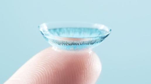
- Soft toric contact lenses: correct regular astigmatism; modern designs stabilize lens axis. Daily disposables reduce infection risk compared with reusable lenses.
- Rigid gas-permeable (RGP) or scleral contact lenses: first-line for irregular astigmatism (eg, keratoconus) to mask corneal irregularity.
- Orthokeratology (overnight corneal reshaping): FDA-cleared devices can reduce low-to-moderate corneal astigmatism in selected corneas; requires adherence and close follow-up; infection risk with overnight wear must be discussed [7].
Adjuncts and special situations
- Manage contributory surface disease (eg, dry eye, blepharitis) to stabilize refraction and contact lens tolerance.
- Cycloplegic refraction in children or when latent hyperopia suspected (eg, cyclopentolate 1% as above; refer to product labeling for age-specific use) [5].
No pharmacologic therapy corrects astigmatism itself.
Procedural and surgical management
- Laser refractive surgery
- LASIK and PRK: excimer laser reshapes cornea to neutralize astigmatism; requires adequate corneal thickness, stable refraction, and absence of ectasia risk factors; FDA-approved platforms treat myopic, hyperopic, and mixed astigmatism within specified ranges [1][8].
- SMILE (small incision lenticule extraction): femtosecond laser–based corneal lenticule removal; FDA-approved for myopia with astigmatism within defined parameters [8].
- Corneal relaxing incisions
- Manual or femtosecond arcuate keratotomy/limbal relaxing incisions to reduce low-to-moderate corneal astigmatism, commonly combined with cataract surgery.
- Cataract surgery with astigmatism management
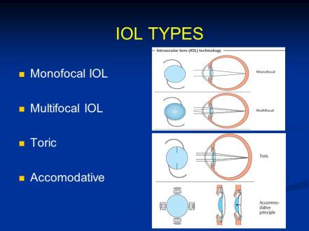
- Toric intraocular lenses (IOLs): align cylinder axis at implantation; effective for regular corneal astigmatism when posterior corneal power is accounted for; rotational stability and posterior corneal astigmatism forecasting are key [9].
- Intraoperative aberrometry/topography and arcuate keratotomy may refine residual astigmatism.
- Irregular astigmatism due to ectasia
- Corneal collagen cross-linking to halt progression in keratoconus/post-ectasia; optical correction with RGP/scleral lenses remains primary for vision [6].
- Intracorneal ring segments as adjuncts in select keratoconus cases to regularize corneal shape.
Patients should receive individualized counseling on risks, benefits, alternatives, and realistic visual expectations. Pregnancy, unstable refraction, and corneal disease are typical contraindications for elective refractive surgery.
Special populations
- Pediatric: Early detection and full optical correction are critical to prevent amblyopia. Refer children with high or asymmetric astigmatism for pediatric ophthalmology assessment and amblyopia management. USPSTF recommends at least one vision screening in children ages 3 to 5 years to detect amblyopia or its risk factors [4].
- Pregnancy/lactation: Corneal curvature and tear film can change; defer refractive surgery during pregnancy and until several months postpartum when refraction is stable. Contact lens tolerance may fluctuate.
- Immunocompromised/contact lens wearers: Emphasize hygiene, avoid overnight wear unless specifically prescribed (eg, orthokeratology under strict protocols), and lower threshold to evaluate for keratitis.
- Elderly: Axis tends to shift toward ATR with age; cataract surgery presents an opportunity to correct astigmatism with toric IOLs or arcuate incisions.
Prognosis
With appropriate optical correction, visual acuity is typically excellent. Children corrected promptly have good amblyopia prevention outcomes; delayed correction increases risk of persistent reduced acuity [3][4]. Irregular astigmatism secondary to ectasia or scarring often requires specialty contact lenses; cross-linking can stabilize progressive ectasia but does not restore normal optics [6]. Surgical correction of regular astigmatism has high patient satisfaction when selection is appropriate and expectations are aligned [1][9].
Prevention and Patient Counseling
- Astigmatism cannot be prevented; routine eye exams detect it early, particularly in childhood.
- Maintain ocular surface health (manage dry eye, blepharitis) to improve visual consistency and contact lens performance.
- For contact lens users: adhere to replacement schedules, avoid sleeping in lenses unless prescribed, and seek care promptly for pain, redness, or light sensitivity to prevent keratitis.
- After cataract or refractive surgery: attend all follow-ups; report sudden blur, pain, or photophobia immediately.
When to Seek Immediate Care
- Sudden vision loss or a dramatic change in vision
- Eye pain, redness, or light sensitivity, especially with contact lenses
- New monocular double vision (double image in one eye) that does not resolve with blinking
- Symptoms after eye injury or after eye surgery (eg, LASIK, PRK, cataract surgery)
- Halos with severe eye pain and nausea
References
- American Academy of Ophthalmology. Refractive Errors & Refractive Surgery Preferred Practice Pattern. 2022. https://www.aao.org/ppp
- National Eye Institute. Facts About Refractive Errors (Astigmatism). https://www.nei.nih.gov/learn-about-eye-health/eye-conditions-and-diseases/refractive-errors
- American Association for Pediatric Ophthalmology and Strabismus (AAPOS). Amblyopia Risk Factors and Treatment Guidelines. 2021. https://aapos.org/glossary/amblyopia
- US Preventive Services Task Force. Vision Screening in Children Aged 3 to 5 Years: Recommendation Statement. JAMA. 2017;318(9):836-844. doi:10.1001/jama.2017.11260
- Cyclopentolate hydrochloride ophthalmic solution prescribing information. DailyMed. https://dailymed.nlm.nih.gov/dailymed/
- EyeWiki. Keratoconus. American Academy of Ophthalmology. https://eyewiki.aao.org/Keratoconus
- Bullimore MA, Johnson LA. Overnight Orthokeratology. Cont Lens Anterior Eye. 2020;43(4):322-332. doi:10.1016/j.clae.2019.10.006
- US Food and Drug Administration. LASIK and SMILE information for patients. https://www.fda.gov/medical-devices/surgery-devices/lasik
- Ferreira TB, et al. Toric intraocular lenses: clinical outcomes and considerations. J Cataract Refract Surg. 2019;45(5):631-648. doi:10.1016/j.jcrs.2019.02.018
Disclaimer: This article is for informational purposes and is not a substitute for professional medical advice, diagnosis, or treatment. Always consult a qualified eye care professional about your specific condition. If you have sudden vision loss, severe eye pain, or an eye injury, seek emergency care immediately.