
What is an intraocular lens (IOL)?
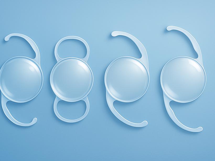
An intraocular lens is a clear, artificial lens placed inside the eye to replace the eye’s cloudy natural lens during cataract surgery. Removing the cloudy lens restores optical clarity, and the IOL restores the eye’s focusing power. The same technology can also be used in refractive lens exchange (RLE), an elective procedure to reduce reliance on glasses or contacts before a visually significant cataract develops. IOLs are designed to be permanent; you do not feel them, and they are not seen by others.
How IOLs improve focus
In a healthy eye, the natural lens bends light so it focuses on the retina. With age, the lens stiffens (presbyopia), reducing near focus, and with cataracts, the lens becomes cloudy, scattering light and degrading contrast. IOLs replace the cloudy lens to restore clarity and, depending on the design chosen, can focus light for one distance or across a range (distance, intermediate, and near). Some IOLs also correct regular corneal astigmatism (uneven corneal curvature that causes blur and ghosting). Each design balances sharpness, range of focus, contrast, and the likelihood of visual phenomena such as halos, glare, or starbursts, which are often more noticeable in dim light.
Main IOL types
Monofocal IOLs
What they do: Provide clear vision at a single focal point—typically set for distance, intermediate (computer), or near (reading) in each eye. Many people choose distance in both eyes and use reading glasses for near tasks.
- Pros: High-quality image and contrast at the chosen distance; generally the lowest likelihood of halos, glare, or starbursts; widely covered by insurance for cataract surgery.
- Cons: Glasses are usually needed for other distances; a single eye has a limited range of focus.
- Variations: Surgeons may use mini-monovision or monovision—setting one eye slightly nearsighted—to reduce dependence on reading glasses. A contact lens trial helps you assess tolerance, as it can modestly affect depth perception.
Enhanced or “extended” monofocal IOLs
What they do: These lenses are still monofocal by regulation but are engineered to provide a slightly broader range than a traditional monofocal—often improving intermediate vision (dashboards or computer) while maintaining distance clarity.
- Pros: Image quality and night-vision profile similar to standard monofocals, with a touch more range; many people use readers only for fine print.
- Cons: Not designed to fully replace reading glasses; the degree of added range varies by design and by individual eye anatomy.
Extended Depth of Focus (EDOF) IOLs
What they do: Create an elongated focal zone to sharpen distance and intermediate vision and offer some functional near. Many patients can use a computer and phone comfortably; fine print may still require readers.
- Pros: Broader range than monofocals; balanced performance for many daily tasks; toric versions are available for regular corneal astigmatism.
- Cons: Some risk of halos, glare, or reduced contrast—typically less than with multifocal designs but still possible, especially at night; night-driving sensitivity varies by person.
Multifocal and trifocal IOLs
What they do: Split light into two or three focal points (distance, intermediate, and near) to reduce dependence on glasses across a wide range.
- Pros: The greatest potential for reduced spectacle dependence across distances; many daily tasks can be performed without glasses.
- Cons: Higher likelihood of halos, glare, starbursts, and reduced contrast sensitivity, especially at night. Neuroadaptation can take weeks to months, and some people remain aware of these effects. Performance can be limited by other eye conditions (such as macular disease, glaucoma that reduces contrast, corneal problems, or significant dry eye). Toric versions exist for regular corneal astigmatism.
- Considerations: People who do a lot of night driving or who are very sensitive to visual artifacts may prefer other options. Careful screening and expectation-setting are important.
Accommodating IOLs
What they do: Designed to shift position or flex slightly inside the eye to provide some range of focus by mimicking natural focusing. In real-world use, the amount of near vision gained is often modest and variable.
- Pros: Potential for improved intermediate vision with a visual profile closer to monofocal for some patients.
- Cons: Near vision improvement may be limited and can change over time; not all surgeons offer these lenses; outcomes can be variable. Glasses are often still needed for fine print.
Toric IOLs (for astigmatism)
What they do: Toric lenses have built-in correction for regular corneal astigmatism and are available as monofocal, enhanced monofocal, EDOF, and multifocal/trifocal designs.
- Pros: Sharper vision by addressing regular corneal astigmatism at the time of surgery; may reduce the need for glasses for the targeted distance(s).
- Cons: Precise alignment is required. If the lens rotates after surgery, vision can blur and a minor repositioning procedure may be needed. Toric IOLs are not designed to correct irregular astigmatism (for example, from keratoconus or corneal scars). For small amounts of regular astigmatism, your surgeon may instead use corneal incisions (manual or femtosecond laser), with or without a toric lens.
Materials and lens design
IOLs are made from medical-grade materials shaped to fit securely in the lens capsule. The material and design influence incision size, optical performance, and how the lens interacts with the eye over time.
- Hydrophobic acrylic: The most common modern material. It is foldable for small incisions and often paired with a sharp posterior edge design that helps limit clouding of the capsule behind the lens. Some older formulations could develop tiny internal reflections (“glistenings”); many newer formulations aim to maintain clarity over time.
- Hydrophilic acrylic (sometimes called hydrogel): Also foldable and used in a variety of situations. Choice depends on surgeon preference and specific eye needs.
- Silicone: Clear and foldable. Usually avoided if silicone oil might be needed in future retinal surgery, as interactions can affect visualization.
- PMMA (polymethyl methacrylate): A rigid plastic used less commonly today because it requires a larger incision, but appropriate in select cases.
- Aspheric optics: Many IOLs incorporate asphericity to reduce spherical aberration and improve image quality, especially in low light. Some lenses are aberration-neutral to better match individual corneas; your surgeon will select the approach that fits your eye’s measurements.
- Haptics and edge design: The supporting arms (haptics) and squared optic edges help stabilize the lens and reduce posterior capsule opacification (a common, treatable cause of blurry vision months or years after surgery).
UV and blue-light filtering
All modern IOLs block ultraviolet (UV) light to protect internal eye structures. Some IOLs also filter a portion of blue light and appear slightly yellow. Blue-light filtering can reduce certain wavelengths that scatter more in the eye and may feel more comfortable to some people. Evidence on whether blue-light filtering prevents retinal disease is mixed. Color perception may feel subtly warmer at first and usually adapts. Your surgeon will consider retinal health, light sensitivity, and personal preference when choosing between clear and blue-filtering designs.
How surgeons match a lens to you
A thorough exam and conversation guide lens choice more than any single feature. Key factors include:
- Precise measurements: Biometry, keratometry, and corneal topography map your eye’s length and curvature. Surface dryness can skew readings, so optimizing the tear film (treating dry eye/blepharitis) before measurements improves accuracy. If you wear contacts, a contact-lens “holiday” (longer for rigid lenses) before biometry helps measurements settle.
- Astigmatism: Amount, orientation, and regularity influence whether a toric IOL or corneal incisions are best. Toric lenses target regular corneal astigmatism; irregular astigmatism may call for different strategies.
- Retina, optic nerve, and cornea health: Conditions like macular degeneration, diabetic retinopathy, glaucoma that reduces contrast, keratoconus/irregular cornea, Fuchs dystrophy, or zonular weakness/pseudoexfoliation may limit benefit from presbyopia-correcting lenses and often favor monofocal strategies.
- Lifestyle and visual priorities: Night driving, computer work, hobbies, and reading needs help determine the balance between range of focus and possible visual effects such as halos or contrast reduction.
- Risk tolerance and adaptation: Some designs trade more range for a higher chance of night-time halos. Neuroadaptation typically takes weeks to months.
- Prior eye surgery: Previous LASIK, PRK, or RK alters measurements and may affect lens calculations and design choice. Surgeons often use additional data or intraoperative tools to refine the target, and small residual refractive errors are more likely.
- Strategy across both eyes: Planning both eyes together—such as blended vision or small offsets—can improve overall function.
- Budget and coverage: Insurance considerations may guide whether premium features are practical.
Costs and insurance in the U.S.
For medically necessary cataract surgery, Medicare and most insurers typically cover a standard monofocal IOL and the procedure. Options that correct astigmatism (toric) or extend range of vision (enhanced monofocal, EDOF, multifocal/trifocal, or accommodating) are usually considered elective upgrades with additional out-of-pocket costs. Refractive lens exchange (RLE) is elective and generally self-pay. Coverage details and fees vary by plan and provider; ask your surgeon’s office for a personalized estimate.
What to expect after surgery
Vision often improves within days, with fine-tuning over several weeks as the eye heals and the brain adapts to the new optics. It is common to notice halos, glare, edge reflections, or the awareness of a “shadow” (negative dysphotopsia) early on; these usually lessen, but a minority of people remain symptomatic. Depending on the IOL and your eye’s healing, you may still use glasses for certain tasks. Small residual refractive errors or astigmatism can sometimes be refined with glasses, contact lenses, corneal incisions, a laser enhancement, or—in selected cases—lens exchange.
If a toric IOL rotates, a straightforward in-office or operating-room repositioning can restore clarity. Months or years later, some people develop posterior capsule opacification (a cloudy membrane behind the IOL) that can be cleared with a brief clinic laser procedure (Nd:YAG capsulotomy). This laser treats the membrane, not optical halos, and once performed it can make later IOL exchange more complex; if you are unhappy with vision quality, discuss options with your surgeon before having a capsulotomy.
Call your surgeon urgently for increasing eye pain, sudden vision loss, worsening redness, light sensitivity, or new flashes/floaters.
Special situations and alternatives
- Small-aperture (“pinhole”) IOLs: In select cases—such as irregular corneas—they can increase depth of focus and mask aberrations, often with some dimming of light. These are niche options.
- Refractive lens exchange (RLE): Can reduce dependence on glasses in presbyopic adults who do not yet have a visually significant cataract. Candidacy is individualized; in some groups (for example, highly myopic patients), RLE can carry a higher risk of retinal detachment compared with waiting for cataract surgery. Discuss your risk profile carefully.
Making your decision
No single IOL is “best” for everyone—each type balances range of focus, image quality, night vision, and cost differently. Discuss your goals, daily tasks, and medical history with your ophthalmologist so you can choose a lens strategy that fits your eyes and your life. Expect some period of adaptation, and remember that even with advanced lenses, complete spectacle independence is not guaranteed.
This article is for general education and is not medical advice. Your optimal IOL choice and surgical plan should be individualized with your ophthalmologist.
Sources
- American Academy of Ophthalmology — IOLs for cataract surgery
- American Academy of Ophthalmology — Presbyopia-correcting IOLs
- National Eye Institute — Cataract surgery